Menu
  • Publish Your Research/Review Articles in our High Quality Journal for just USD $99*+Taxes( *T&C Apply)

    Offer Ends On

Research Article

Sternberg's Canal Defect and It’s Sequelae of Csf Leak, Spontaneous Pneumocephalus and DEJAVU Seizure

Zainab Sachit Hashim*, Ali Saud, Ammar Abdulkadhm, and Hani Musa Bader

Corresponding Author: Zainab Sachit Hashim, Endoscopic Skull Base Surgery Centre, Medical City Teaching Hospital, Baghdad, Iraq.

Received: April 26, 2022 ;    Revised: May 05, 2022 ;    Accepted: May 08, 2022

Citation: Hashim ZS, Saud A, Abdulkadhm A & Bader HM. (2022) Sternberg's Canal Defect and It’s Sequelae of Csf Leak, Spontaneous Pneumocephalus and DEJAVU Seizure. Int J Med Microbiol Immunol

Copyrights: ©2022 Hashim ZS, Saud A, Abdulkadhm A & Bader HM. This is an open-access article distributed under the terms of the Creative Commons Attribution License, which permits unrestricted use, distribution, and reproduction in any medium, provided the original author and source are credited

Share Your Publication :

Views & Citations

1798

Likes & Shares

0


Global Views

  • Abstract
  • Full Text
  • Images
  • Tables
  • References
  • PDF
  • Supplementary Files

Abstract

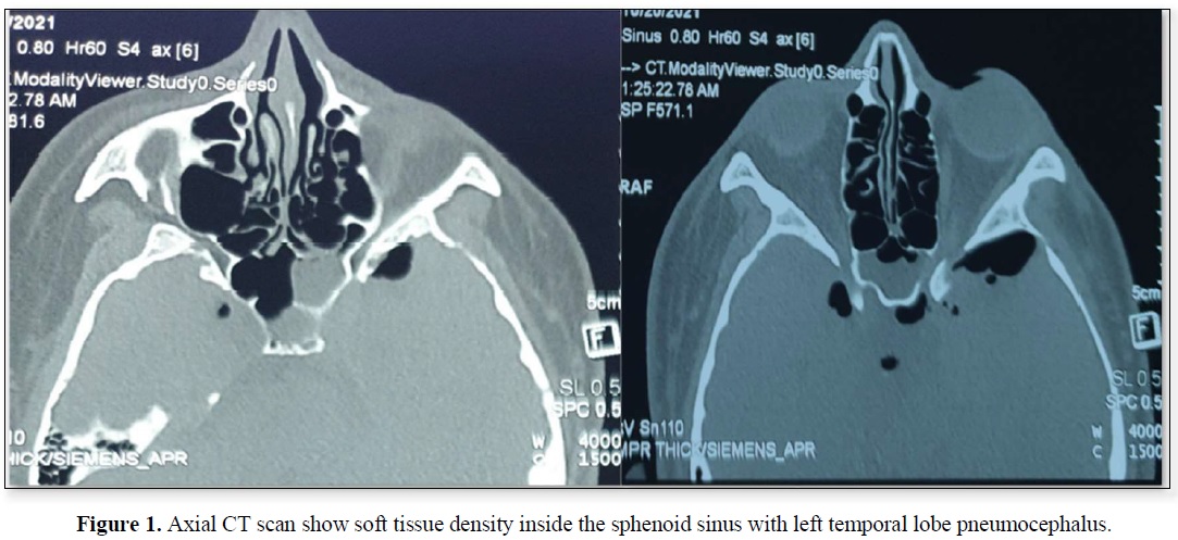
Background: We described a patient with spontaneous pneumocephalus probably arising from Sternberg’s canal of the sphenoid sinus. Spontaneous Pneumocephalus (PNC) associated with Cerebrospinal (CSF) fluid leaks and Intrasphenoidal Meningocephalocele are extremely rare findings and represent a condition bearing serious risks for the patient. Sternberg’s canal is a lateral craniopharyngeal canal resulting from incomplete fusion of the greater wings of the sphenoid bone with the basisphenoid. It acts as a weak spot of the skull base, which may lead to develop a temporal lobe meningocephalocele protruding into the lateral recess of the sphenoid sinus (SS).

Case description: A 27-year-old male, presented with left cerebrospinal fluid (CSF) rhinorrhea, referred from the neurosurgical department with severe headache, and low-grade fever. Radiological investigations consisted of computed Tomography (CT) scan, this imaging study identified a soft tissue density Through a bony defect which communicates the middle cranial fossa with the lateral recess of the SS, and pneumocephalus of left temporal lobe.  Patient underwent an Endoscopic Endonasal repair of the defect. There was no complication related to the surgical procedure and no recurrence of CSF leakage occurred 4 months after surgery.

Conclusion: A persisting Sternberg’s canal should be considered the source of Spontaneous Pneumocephalus and CSF-leaks with or without meningoencephaloceles in sphenoid sinuses with extensive lateral pneumatization, especially when located laterally and below the maxillary nerve. Endoscopic endonasal surgery is safe as no intraoperative complications occurred in our patient. It is less traumatic, providing a good access and view of the surgical field. Our case is among the rare reported cases.

Keywords: Cerebrospinal fluid rhinorrhea, Intrasphenoidal meningoencephalocele, Lateral craniopharyngeal canal, Lateral recess, Sphenoid sinus, Sternberg’s canal

INTRODUCTION

Pneumocephalus is defined as intracranial air (Extracerebral, Intracerebral, Intravascular compartment), result from an abnormal communication between an air-containing structure (most commonly the paranasal sinuses and temporal bones) and the subarachnoid space through a tear in the meninges and a fracture of the skull base. The majority of pneumocephalus cases are due to trauma. Spontaneous pneumocephalus is a rare condition, which represented only 0.6% of pneumocephalus occur due to osteodural defect adjacent to the paranasal sinuses/Sternberg’s canal. tension pneumocephalus, air under pressure, leading to serious neurological complications or even death. Such cases call for urgent repair of the Skull Base defect regardless of its etiology decreases by 10-15 days and never is present by 3 weeks [1-3].

The sphenoid sinus spontaneous leaks which; although rare, are more common in this location than secondary leaks. Embryologically, the sphenoid bone develops from four parts: the presphenoid (forming the anterior sphenoid bone; the basisphenoid (forming the posterior sphenoid bone); the orbitosphenoid (forming lesser wings); and the alisphenoid (forming greater wings and lateral parts of the pterygoid process). The fusion of which occurs after birth from the anterior to posterior regions. Just before complete fusion, a canal-connecting middle cranial fossa and nasopharynx called the lateral craniophangeal" or Sternberg's canal (named after Sternberg in 1888, although first described by in 1877) is formed. This canal closes by the age of ten years through ossification. Persistence of this canal into adulthood may be the cause of a potential CSF leak. persistent Sternberg's canal incidence ranges from 0.42-6.19%) [4].

CASE PRESENTATION

Case 1

A 27-year-old male, pharmacist, married, have no child, known case of tonic-clonic focal seizure (Déjà vu) with chronic use of keppra 500 mg {1.5 g × 2} since 2018, Referred from neurologist to the Neuro rhinology/ENT department. As a history of 3-days unilateral left sided clear, watery rhinorrhea, as drops then became profuse amount salty in taste preceded by one attack of (Déjà vu) at a period of less than 1 week, associated with severe headache aggravated by head movement (increase in supine, decrease in sitting position) with drowsiness, left sided pulsatile tinnitus and low-grade fever.

Physical examination revealed left-sided rhinorrhea. The nasal discharge was interpreted as cerebrospinal fluid (CSF) rhinorrhea.

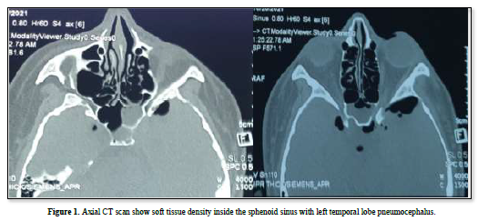
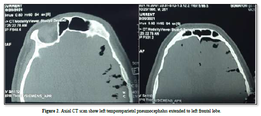
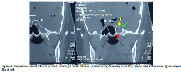
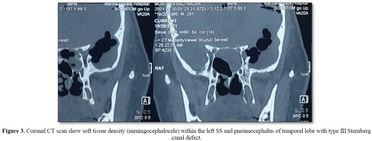
A computed tomography (CT) scan revealed a defect in the middle cranial fossa communicated to the lateral recess of the left Sphenoid Sinus and a soft tissue density within the sinus, with air void signal extended in to the left temporoparietal and frontal hemisphere (Figures 1-4). It appeared to be due to persistence of the Sternberg’s canal.

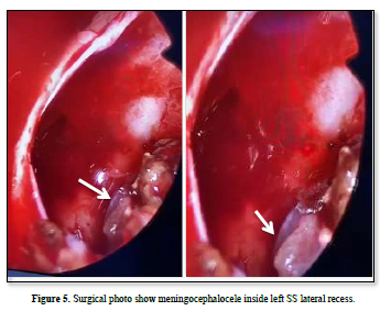
The patient underwent endoscopic endonasal trans-sphenoidal approach surgery by a Neurorhinologist/ Endoscopic Skull base surgeon. The bony defect in the Left SS lateral recess was identified during the procedure and the protruded meningencephaloocele (Figure 5) was reduced in volume by bipolar cauterization and the defect was filled with bone of middle turbinate, fat and fascia lata from anterolateral aspect of thigh.

There was no complication related to the surgical procedure. No recurrence of CSF leakage occurred after 4 months of surgery.

DISCUSSION

  • Spontaneous, nontraumatic pneumocephalus is very uncommon, and most cases result from nose blowing, sneezing, and the valsalva maneuver and by environmental pressures including those encountered in mountain climbing, during flights, and during scuba diving [8]. The presenting symptom in pneumocephalus is usually Headache, and other symptoms include CSF rhinorrhea (as in our patient), meningeal signs, hemiparesis, papilledema, and cranial nerve palsies. However, the presentation of pneumocephalus (PNC) is often vague.
  • Only 3 cases of spontaneous nontraumatic pneumocephalus (PNC) are associated with lateral recess defect (one in 1992 and other in 1999), new reported case (one in 2010) [5-7].
  • Intrasphenoidal temporal lobe encephalocele protruding in the Lateral sphenoid sinus is one of the most unusual types of basal encephalocele (as in our case). previous reports have shown that temporal lobe encephaloceles in the lateral sphenoid sinus present with a variety of signs and symptoms, such as CSF rhinorrhea, meningitis, headache, vertigo, seizure, and some somatic complaints [7].
  • Temporal lobe seizures begin in the temporal lobes of brain, which process emotions and are important for short-term memory. Some symptoms of a temporal lobe seizure may be related to these functions, including having odd feelings - such as euphoria, deja vu or fear. Temporal lobe seizures are sometimes called focal seizures with impaired awareness. Some people remain aware of what's happening, but during more-intense seizures, you might look awake but be unresponsive. Temporal lobe seizures may stem from an anatomical defect (as in our patient) or scar in temporal lobe, but the cause is often unknown [9].
  • Spontaneous CSF-leaks occur without any history of trauma, iatrogenic injury, tumor, malformations or radiotherapy [10]. Some of those leaks originate from a bony dehiscence in the lateral sphenoid sinus wall called Sternberg’s canal (as in our patient). This special entity of spontaneous CSF-leaks is a rare finding. Castelnuovo [11] reported 15 patients with a CSF-leak originating from Sternberg’s canal. Blaivie [12] and Schick [13] reported one case each with the same pathology. Our study includes one case also.
  • Obesity is thought to be a risk factor for spontaneous CSF leak and meningoencephalocele (as in our patient). The theoretic physiopathology of its association is that increased weight increases intraabdominal and intrathoracic pressure which could lead to the development of benign intracranial hypertension [14].
  • Radiological images, CT scan is a noninvasive imaging technique which gives good bone detail and identifies the site of the skull base defect and may also show partial or complete opacity of the SS [15]. MR images give better information about the soft tissues like the meningocele itself [16]. Our patient's imaging was showed meningoencephalocele, pneumocephalus and lateral recess defect.
  • The diagnosis is confirmed at the time of surgery [17]. Persistent CSF leak is potentially lethal because it may lead to meningitis or brain abscess. Thus, repair of intrasphenoidal meningoencephalocele has two main objectives: prevention of CSF leak and to avoid central nervous infection.
  • Surgical treatment should be tailored to each patient. Endoscopic Trans nasal Approaches are less invasive and do not require a large external incision and temporal lobe retraction, minimizing brain manipulation [18]. Our patient underwent Endoscopic Endnasal surgery for repairing successfully.

CONCLUSION

  • A persisting Sternberg’s canal should be considered the source of spontaneous pneumocephalus and CSF-leaks with or without meningoencephaloceles in sphenoid sinuses with extensive lateral pneumatization, especially when located laterally and below the maxillary nerve.
  • Endoscopic endonasal surgery is safe as no intraoperative complications occurred in our patient. It is less traumatic, providing a good access and view of the surgical field.

Our case is among the very uncommon cases of spontaneous pneumocephalus in lateral recess defect of sphenoid sinus.

REFERENCES

  1. Acton QA (2012) Issues in CNS Diseases and Disorders: 2011 Edition. By scholarly editions, Atlanta, Georgia.
  2. Khanna A (2019) Cerebrospinal Fluid Rhinorrhea and Spontaneous Pneumocephalus in a Patient with Inactive Granulomatosis with Polyangiitis. Morressier.
  3. Jhaveri MD, Salzman KL, Ross JS, Osborn JS, Ho CY (2018) ExpertDDx: Brain and Spine, 2nd Philadelphia, Elsevier.
  4. Hegde JS, Vamanshankar H (2020) CSF Rhinorrhea Management and Practice, 1st, Boca Raton, CRC Press.
  5. Cartwright MJ, Eisenberg MB (1992) Tension pneumocephalus associated with rupture of a middle fossa encephalocele. Case report. J Neurosurg 76(2): 292-295.
  6. Shafa B, Arle J, Kotapka M (1999) Unusual presentations of middle fossa encephaloceles: Report of two cases. Skull Base Surg 9(4): 289-294.
  7. Ohkawa T, Nakao N, Uematsu Y, Itakura T (2010) Temporal lobe encephalocele in the lateral recess of the sphenoid sinus presenting with intraventricular tension pneumocephalus. Skull Base 20(6): 481-486.
  8. Baba M, Tarar O, Syed A (2016) A Rare Case of Spontaneous Pneumocephalus Associated with Nontraumatic Cerebrospinal Fluid Leak. Case Rep Neurologic Med 2016: 1828461.
  9. Temporal lobe seizure. Available online at: https://www.mayoclinic.org/diseases-conditions/temporal-lobe seizure/symptoms-causes/syc-20378214
  10. Schuknecht B, Simmen D, Briner HR, Holzmann D (2008) Nontraumatic Skull Base Defects with Spontaneous CSF Rhinorrhea and Arachnoid Herniation: Imaging Findings and Correlation with Endoscopic Sinus Surgery in 27 Patients. AJNR 29: 542-549.
  11. Castelnuovo P, Dallan I, Pistochini A, Battaglia P, Locatelli D, et al. (2007) Endonasal endoscopic repair of Sternberg’s canal cerebrospinal fluid leaks. Laryngoscope 117: 345-349.
  12. Blaivie C, Lequeux T, Kampouridis S, Louryan S, Saussez S (2006) Congenital transsphenoidal meningocele: Case report and review of the literature. Am J Otolaryngol 27: 422-444.
  13. Schick B, Brors D, Prescher A (2000) Sternberg’s canal - cause of congenital sphenoidal meningocele. Eur Arch Otorhinolaryngol 257: 430-432.
  14. Lai SY, Kennedy DW, Bolger WE (2002) Sphenoid encephaloceles: Disease management and identification of lesions within the lateral recess of the sphenoid sinus. Laryngoscope 112: 1800-1805.
  15. Martin TJ, Smith TL, Smith MM, Loehrl TA (2002) Evaluation and surgical management of isolated sphenoid sinus disease. Arch Otolaryngol Head Neck Surg 128: 1413-1419.
  16. Conboy PJ, Johnson IJ, Jaspan T, Jones NS (1998) Idiopathic diffuse erosion of the skull base presenting as cerebrospinal fluid rhinorrhea. J Laryngol Otol 112: 679-681.
  17. Tabaee A, Anand VK, Cappabianca P, Stamm A, Esposito F, et al. (2010) Endoscopic management of spontaneous meningoencephalocele of the lateral sphenoid sinus. J Neurosurg 112: 1070-1077.
  18. Castelnuovo P, Dallan I, Pistochini A, Battaglia P, Locatelli D, et al. (2007) Endonasal endoscopic repair of Sternberg’s canal cerebrospinal fluid leaks. Laryngoscope 117: 345-349.

    No Files Found