
-
Publish Your Research/Review Articles in our High Quality Journal for just USD $99*+Taxes( *T&C Apply)
Offer Ends On
Publish Your Research/Review Articles in our High Quality Journal for just USD $99*+Taxes( *T&C Apply)
Offer Ends On
Rupali R Taur* and Shantanu Y Chavan
Corresponding Author: Rupali R Taur, Department of Biosciences and Technology, MGM University, Chhatrapati Sambhajinagar 431003 (MS) India.
Received: February 21, 2025 ; Revised: March 04, 2025 ; Accepted: March 07, 2025 ; Available Online: April 09, 2025
Citation: Taur RR & Chavan SY. (2025) Green Synthesis of Silver Nanoparticles Using the Leaf Extract of Withania somnifera (L.) Dunal. J Pharm Sci Drug Discov, 4(1): 1-8.
Copyrights: ©2025 Taur RR & Chavan SY. This is an open-access article distributed under the terms of the Creative Commons Attribution License, which permits unrestricted use, distribution, and reproduction in any medium, provided the original author and source are credited.
Views & Citations
Likes & Shares
Presently, research on the synthesis of noble metal nanoparticles (NPs) has advanced and attracted significant attention. The use of plant extracts is the selected procedure for the biological synthesis of NPs due to the presence of biologically valuable compounds. Withania somnifera is a plant conferred with medicinal benefits in specifically Alzheimer's disease and Parkinson's disease due to its exclusive curative properties. The main objective of this study is too the rapid and simple and efficient synthesis of silver nanoparticles by Withania somnifera Linn. The exposure of reaction mixtures of fresh leaf extract of W. somnifera with silver nitrate derived in reduction of metal ions within10 min. The extracellular synthesized silver nanoparticles were characterized by scanning electron microscopy, X-ray diffraction studies, and Fourier transform IR. From the results, W. somnifera sliver nanoparticle AgNPs revealed that significantly higher concentration of AgNPs. The key findings of this investigation shown that synthesized Ag-NPs demonstrate significant medicinal activity and could be potentially ideal alternatives in medicinal applications.
Keywords: Silver nanoparticles, Medicinal activity, Withania somnifera, Molecular characterizations
INTRODUCTION
Nanotechnology involves the synthesis of nanoparticles (NPs) with various morphology, size, and to be used for their application in different Fields. Green synthesis mode using many plants parts have developed active materials that have significant power against gram-positive and gram-negative microorganisms, many cancer cell lines and presence of higher antioxidant effects than those produced by chemical and thermal physical methods [1]. The various plant products, the leaves of plants have gained much attention for application. The synthesis of biomedically important NPs (nanoparticles) using leaf extracts has various applications compared with other chemical methods [2]. Because of their specific chemical properties.
In current, the silver nanoparticles (AgNPs) have received a significant interest from many researchers and scientist those working on different disciplines due to their specific features and a broad spectrum of implementation [3] for occurrence, in food science, medical, agriculture, and agricultural technologies [4] Previous studies have reported that AgNPs have significant antimicrobial activities against Escherichia coli, Staphylococcus aureus, and Serratia marcescens [5]. It also reported that antinematodes, anti-viral anti-cancer and anti-inflammatory effects [6-9]. Ashwagandha (Withania somnifera, fam. Solanaceae) is widely known as “Indian Winter cherry” or “Indian Ginseng”. It is one of the most valuable herbs of Ayurveda (the traditional system of medicine in India) used for millennia as a Rasayana for its broad ranging health therapeutic. Rasayana is described as an herbal or metallic preparation that enhances a youthful state of physical and mental health and increases happiness [10]. Types of therapy are given to small children as tonics, and are also given by the middle-aged and elderly to enhance longevity. Among the ayurvedic Rasayana herbs, it was also exhibit useful in neurodegenerative diseases such as Parkinson’s, Huntington’s and Alzeimer’s diseases.
Application of biosynthesized AgNPs is significant interest in the field of agriculture because of their antioxidant and wide spectrum of antimicrobial activity along with their eco-friendly, biocompatible, and cost-effective nature. However, of keen interest to optimize the inhibitory effect of synthesized AgNPs against antioxidant and antibacterial activity which can be implemented in the field of nanotechnology as a cost-efficient, environmentally friendly and safe strategy. Therefore, the objective of current study was to synthesize SNPs using fresh leaf extract of W. somnifera and their presence confirmed with SEM-EDAX and FTIR characterization.
MATERIALS AND METHODS
Extract preparation
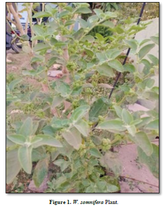
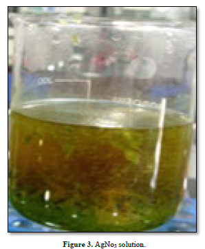
Fresh leaves of Withania somnifera were collected from Dr. Babasaheb Ambedkar Matathwada University, Botanical garden Aurangabad (Figures 1 & 2). The fresh leaf of Withania somnifera extract was prepared by taking 10 gm of thoroughly washed and finely cut leaves in 250 ml conical flask along with 100 ml of sterilized glass distilled water and then boiling mixture for 5 min before finally decanting it. At room temperature (Figure 3), 5 mM aqueous silver nitrate solution was added (Figure 4). The extract was filtered through Whatman’s filter paper No. 01. This filtered extract was stored at 4°C in the refrigerator for further studies and analysis.

NANOPARTICLES SYNTHESIS AND CHARACTERIZATION
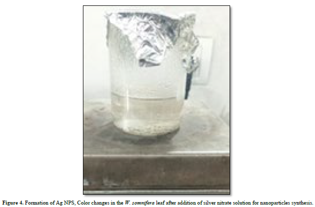
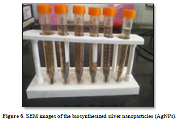
The initial time of color change of reaction mixture was evaluated by visual observation (Figures 5 & 6), soon after the green synthesis the An-AgNPs were evaluated in Nanodrop-8000, UV-visible spectrometer, and recorded its surface plasmon resonance (SPR) peak. Fourier transform infra-red spectroscopy (FTIR) analysis of Withania somnifera leaf extract and green synthesized AgNPs (Figures 7 & 8) was carried out (Bruker Tensor 27) to find study the morphology in the leaf extract which are actively involved in bio-reduction and green synthesis of AgNPs.

Scanning Electron microscopy (SEM) and EDAX analysis was done by using (SU8010, Hitachi) machine, used to characterize the shape of AgNPs. On the carbon-coated copper grid, a thin film of the dried samples was made by simply placing a little sample followed by drying for 10 min in the presence of mercury lamp. A Field-Emission Scanning Electron Microscope (FESEM) images were used to study the size and morphology of AgNPs
RESULTS
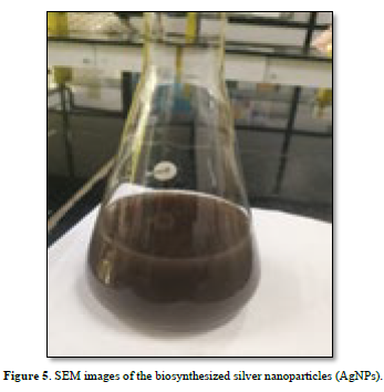
In the present investigation, these AgNPs were synthesized by using aqueous leaves extracts of Withania somnifera by incubating with silver nitrate. A color change of the medium from faint to dark brown color was developed by the addition of silver nitrate. The appearances of dark brown Color indicated the presence of SNP (Figure 5).
CONFIRMATION by FTIR
The plant crude extract was used to produce the nanoparticles and the synthesized nanoparticles was analyzed with FTIR for identification of functional groups of compounds. The FTIR capacity was similar to conclude the biomolecules that specially bound on the silver particles which are involved in the reduction, capping, and stabilization. FTIR spectra of W. somnifera leaf extract showed sharp absorption peaks at 3514/cm, 3859/cm, 1539/cm, 925/cm, 816/cm, 510/cm, 1068/cm, (Figure 9) The spectrum graphs of sample revealed the variation in peak areas which profound the formation of nanoparticle.
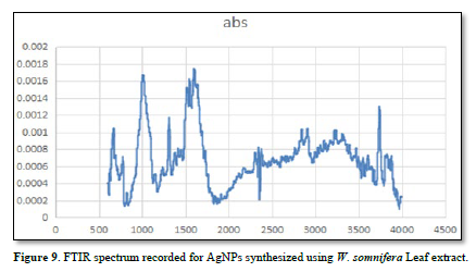
The molecules consisting of aliphatic or aromatic nitro groups, N-H bend of amides or amines, the presence of CH3 and CH2 bending vibrations were observed in the area of 1539/cm. Similarly, the stretching and bending vibrations of C=C and N-H groups from aromatic amines observe the peak at 1539/cm. The sample peak at 2914 and 3890/cm is reported to OH stretching in alcohols and phenolics compounds, 1539/cm reveals the C=H stretch and presence of alkenes groups. The absorption peak present at 1539/cm proteins are interacting with nanoparticles and its secondary structures were not have significant effect during reaction with silver ions [11].
SCANNING ELECTRON MICROSCOPY (SEM)
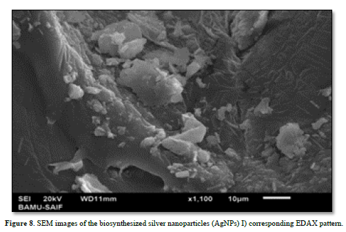
Scanning Electron Microscopy was performed to confirm the development of silver nanostructure. The control sample shows high density silver nanoparticles of Withania somnifera (Figure 8). SEM micrograph analysis finally revealed the synthesis of silver nanoparticles in the reaction mixture. At room temperature, the addition of 5 mM aqueous silver nitrate solution to the leaf extract resulted in the precipitation of nanoparticle. These precipitates are analyzed under microscopic studies for the nanoparticles surface morphology. SEM analysis aqueous leaf extract exhibited spherical and cubical shape. It also observed that silver nanoparticles found with different sizes of the particles. It is concluded from the SEM pictures that silver nanoparticles in the bio reduced colloidal suspensions was measured 59-110 nm in size. The morphology of the nanoparticles was spherical and cubical (Figure 8).
EDX ANALYSIS
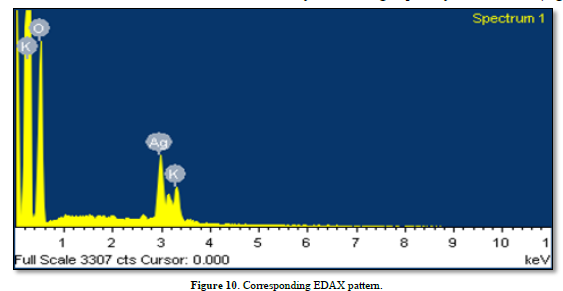
The EDX signals revealed the presence of the silver element in the synthesized AgNPs with a peak optical absorption range, which has been observed in the biosynthesized AgNPs using W. somnifera EDX peaks from, O, K and Ag may show signal is due to the carbon coated copper grid or by the emission of X- rays from proteins and enzymes of leaf extract while the nanoparticles can be attached to either by free amino groups or cysteine residues (Figure 10).
DISCUSSION
The formation of W. somnifera silver nanoparticles by using leaf aqueous extracts is confirmed by formation of dark brown color led to reduction of silver nitrate which indicates the presence of electron donor group in aqueous extract of fresh leaf. The color change in the solution is due to the surface-Plasmon resonance (SPR) phenomena and the dark brown color formation is because of excitation of surface plasmon vibrations in silver nanoparticles. Our results are similar to [11]. It is well known that biosynthesis of nanoparticles application of plant extract is significant strategy for biosynthesis reaction because of their nontoxic ability and thus results in natural capping agent. Our findings here described that synthesis of AgNPs presented after displaying the silver nitrate to W. somnifera leaf extract. The change in dark brown color was observed due to the succession of nanoparticles formation, which has been later analyzed by ultraviolet visible spectroscopy. Biosynthesized AgNPs had a strong band of absorption at 410 nm, resulted in to its SRP attributes. Here, the intensity observed of the color change from light yellowish to dark brown is directly related to the quantity of the extract and incubation period, and this is preferably because of the lasmon vibrations and AgNO3 reduction [12] FTIR studies have been carried out. In case of AgNPs, a shift in the absorbance peak with variation band intensity was found at different regions. AgNPs spectra revealed different absorption bands ranging from 508 to 3914 cm-1. This indicates the presence of possible biomolecules that are involved in reduction and stabilization of silver ions (AgC) to AgNPs (Ag0) which are present in aqueous leaf extract. In Similar with previous report, the FTIR spectrum analysis in this study detected several absorption peaks, specifically for N-H stretching vibrations (Figure 9) indicating strong hydrogen bonding, and C = O extension vibrations attributed to carboxylic acids, ketones, and aldehydes, which were linked to the silver ions reduction resulting to nanoparticles stabilization because of oxidizing the hydroxyl radical [13]. However, the respective SEM-EDAX pattern of the synthesized AgNPs demonstrated the crystalline structure of the particles, which was again confirmed by the energy dispersive X-ray analysis (EDX) results obtained. The EDX signals confirmed an existence of the silver element in the synthesized AgNPs with obtained peak of optical absorption range, which has been observed in the biosynthesized AgNPs using M. capitata [14] and A. nervosa [15] and W. somnifera [16] leaf extract.
Metallic silver nanocrystals generally show typical EDX signals, which is typical for the absorption of metallic silver [17] EDX peaks from C, O, and Cl may be caused due to the carbon coated copper grid or by the emission of X- rays from proteins and enzymes of fruit extract while the nanoparticles can be attached to either by free amino groups or cysteine residues [18].
In conclusion, this study clearly indicates a commercial, environmentally friendly, and reproducible approach in AgNPs synthesis using W. somnifera leaf extracts as a reducing, stabilizing, and capping agent. however, there is extensive research scope in this promising and challenging field. Thus, it is the best platform deserving further exploration for clinical applications (Figure 10).
CONCLUSION
In conclusion, this present study stated that green mediated synthesis of silver nanoparticles using leaf extract of W. somnifera is a novel, simple, cost effective and rapid method for extracellular synthesis of metallic silver nanoparticles. The FTIR analysis shown that broad peak present at 450 nm for silver nanoparticles. Thus, the synthesized nanoparticles showed cuboidal abd spherical structures. Presence of compounds and purity was confirmed by EDAX analysis. The present procedure may use as in the pharmacy industry. Hence, the biological approach appears to deserving exploration in medical application to treat various diseases.
SOURCE OF SUPPORT
The author(s) received no financial support for the research, authorship, and/or publication of this article.
CONFLICT OF INTEREST
The author(s) declared no potential conflicts of interest with respect to the research, authorship, and/or publication of this article.


No Files Found


Internationally Accepted
Share Your Publication :