
-
Publish Your Research/Review Articles in our High Quality Journal for just USD $99*+Taxes( *T&C Apply)
Offer Ends On
Publish Your Research/Review Articles in our High Quality Journal for just USD $99*+Taxes( *T&C Apply)
Offer Ends On
Soni Patel, Nilotpol Kashyap*, Shreya Rani and Jayshree Kalita
Corresponding Author: Nilotpol Kashyap, Department of Pediatric & Preventive Dentistry Vananchal Dental College, Garhwa, Jharkhand, India.
Received: August 09, 2022 ; Revised: September 05, 2022 ; Accepted: September 08, 2022 ; Available Online: September 16, 2022
Citation: Patel S, Kashyap N, Rani S & Kalita J. (2022) A Case Report on Two Cases of Radix Entomolaris in Primary Teeth. J Oral Health Dent Res, 2(2): 1-3.
Copyrights: ©2022 Patel S, Kashyap N, Rani S & Kalita J. This is an open-access article distributed under the terms of the Creative Commons Attribution License, which permits unrestricted use, distribution, and reproduction in any medium, provided the original author and source are credited.
Views & Citations
Likes & Shares
Abstract
Pediatric Dentistry is not only about the hard and soft tissues which are visible and can be treated. There are many dental anomalies which cannot be seen through naked eyes in spite of them being in the oral cavity i.e., anomalies of the root.
These anomalies are detected only when patients undergo treatment for the culprit tooth and these are generally seen in the tooth which undergoes radiographic examination for diagnosis.
During diagnosis primary molar radiograph shows extra root (Radix Entomolaris & Paramolaris). The normal anatomy of mandibular molars consists of two roots but in rarest of the rare conditions there are three roots. When this extra root is present distolingually to the main distal root it is called “Radix Entomolaris” and when it is present mesiobuccally to main mesial root it is called “Paramolaris”. The purpose of this article is to discuss the prevalence, anatomy, classification, clinical and radiographic diagnosis and significance of supernumerary roots in contemporary clinical pediatric dental practice.
Keywords: Radix entomolaris, Radix paramolaris periapical radiographs
INTRODUCTION
The morphology of primary teeth in comparison to permanent is different in its enamel & dentin thickness, more accentuated pulp horns which lead to early spread of infection to the pulp once caries starts. Pediatric endodontics is the method to remove infection from the coronal and radicular pulp followed by chemico-mechanical cleaning and obturating with suitable material. Generally mandibular molars (primary or permanent dentition) have two roots that is mesial and distal with three canals and sometimes four canals which is also very rare. Two canals in the mesial root being mesiobuccal and mesiolingual canals and one canal in distal root [1-3].
Sometimes two canals are also present in distal canal which often creates a rare chance of presence of extra root termed as “Radix entomolaris” or “Paramolaris” [4].
The occurrence of an extra distal root in these mandibular molars is considered as a racial characteristic of certain native Indian and mongoloid populations.
This extra root should also be endodontically treated and obuturated with suitable obturating material to prevent premature extraction.
CASE REPORT
Case 1
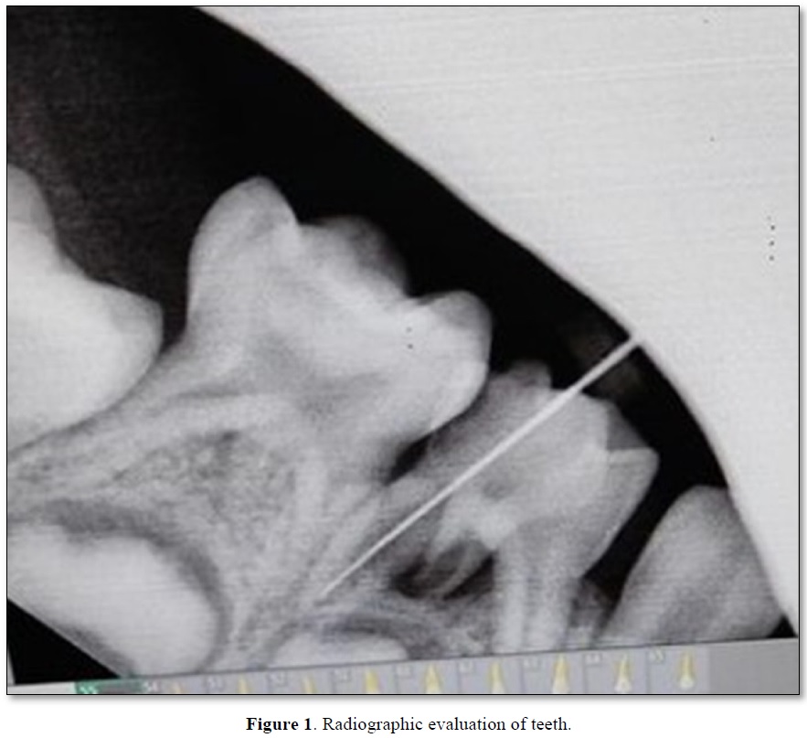
A 5 years old male patient came to the department of pedodontics and preventive dentistry in Vananchal Dental College and Hospital, Garhwa with a chief complain of pain and swelling in lower right back tooth region since 4 to 5 days. Access opening was done followed by working length determination with No.10K file and RVG was taken. On Radiographic evaluation an extra root was detected (Figure 1). Chemico-mechanical preparation was done and then patient was recalled after 2 days.
After two days the pain and swelling had not subsided so the tooth was planned for extraction. The primary molar was thus extracted.
Case 2
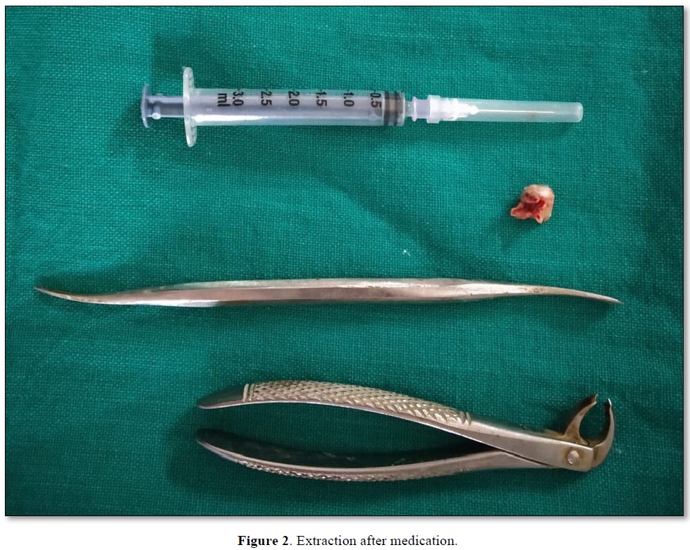
A 10 years old male patient came to the department with a chief complain of pain, swelling and mobility in lower right back tooth region. Medication was given and the patient was recalled after 3 days for extraction as on radiographic evaluation it was found that more than half of the roots were resorbed and interradicular radiolucency was also present.
After extraction it was observed that the mandibular primary molar had three roots (Figure 2).


DISCUSSION
The success of pediatric endodontics is determined by thorough clinical examination, diagnosis, adequate chemo-mechanical preparation, and three-dimensional obturation with suitable obturating material [5].
The first stage of endodontic triad, i.e., correct diagnosis is one of the most important steps towards the success of the endodontic procedure. One of the main reasons for the failure of root canal treatment is incomplete removal of pulpal tissue and microbes from all the pulp canals. Hence, proper radiographic diagnosis plays a very important role in the successful treatment of endodontic therapy to rule out presence of any extra roots [6].
So, radiographs must be taken at different angulations to decrease the chances of “missed canals”. The prevalence rate of radix entomolaris is less than 5% in the Indian population and such cases are not seen commonly during dental treatment. The exact etiology of radix entomolaris is still idiopatheic but according to some authors it may be due to disturbance during odontogenesis or may be due to the high degree of genetic penetrance [7].
To avoid the failure of endodontic therapy, minimum of two different angulated diagnostic radiographs must be taken with careful clinical examination. If radix entomolaris is diagnosed in the radiograph before commencing the endodontic treatment, the access cavity design should be modified trapezoidal so that the additional canal orifice can be easily accessed [8,9].
Through the knowledge of the law of symmetry & various methods like visualization of the dentinal map and bleeding points in the canal orifice, using magnification, ultrasonic tips, staining of the pulp chamber floor with 1% methylene blue dye, performing champagne bubble test, and cone beam computed tomography imaging extra roots and canals can be identified.
According to De Moor [10] the morphology of radix entomolaris canals in the majority of cases were curved. Hence after initial root canal exploration with small files (size 10 or less) together with radiographic working length and curvature determination, the creation of straight-line access and preparation of glide path has to be emphasized to avoid procedural errors.
CONCLUSION
The presence of extra root and its complex anatomical morphology creates a challenge for endodontic therapy. Prognosis of endodontic therapy is determined by the identification of radix entomolaris and paramolaris. A 30º mesial and distal angulations preoperative radiographs and their proper interpretation are mandatory to prevent any misdiagnosis of radix entomolaris. Hence accurate radiographic diagnosis, thorough knowledge about the variation in root canal morphology, prevalence and canal configuration of radix entomolaris are the major factors for the success of endodontic treatment.

REFERENCES
No Files Found


Internationally Accepted
Share Your Publication :