
-
Publish Your Research/Review Articles in our High Quality Journal for just USD $99*+Taxes( *T&C Apply)
Offer Ends On
Publish Your Research/Review Articles in our High Quality Journal for just USD $99*+Taxes( *T&C Apply)
Offer Ends On
Alven Arreaza*, MSc, PhD
Corresponding Author: Alven Arreaza, Oral Medicine - Private Practice, Caracas, Venezuela, 1050 - El Recreo, Alto Centro Building, Apt. 6H.
Received: September 02, 2026 ; Revised: September 04, 2026 ; Accepted: September 06, 2026 ; Available Online: September 08, 2026
Citation: Arreaza A. (2026) Carcinoma of the Mandibular Alveolar Ridge: Case Report and Discussion of a Non-Surgical Approach. J Oral Health Dent Res, 5(3): 1-5.
Copyrights: ©2026 Arreaza A. This is an open-access article distributed under the terms of the Creative Commons Attribution License, which permits unrestricted use, distribution, and reproduction in any medium, provided the original author and source are credited.
Views & Citations
Likes & Shares
A 66-year-old male presented with a painful, bleeding lesion of the lower mandibular alveolar ridge, developing nine months after a tooth extraction. After multiple unsuccessful courses of antibiotics, clinical, imaging, and histopathological evaluation confirmed a well-differentiated squamous cell carcinoma with areas of dedifferentiation. Bone involvement allowed classification as T4a. Due to the expected functional impact of mandibulectomy, concomitant chemoradiotherapy with curative intent was selected. This case illustrates the complexity of therapeutic decision-making in locally advanced oral carcinomas.
Keywords: Oral squamous cell carcinoma; oral cancer; chemoradiotherapy; alveolar ridge; case report.
INTRODUCTION
Head and neck squamous cell carcinoma (HNSCC), particularly that arising in the oral cavity, accounts for more than 90% of malignant neoplasms in this anatomical region and represents a therapeutic challenge due to its biological aggressiveness and proximity to vital osseous structures (1). Traditionally, the standard treatment for advanced stages or tumors with suspected bone invasion has been radical surgical resection with wide oncologic margins. In cases with mandibular involvement, ablative procedures such as marginal or segmental mandibulectomy entail markedly mutilating consequences (2). These interventions produce significant facial deformities and severe functional impairment, critically affecting swallowing, mastication, and speech, often resulting in profound psychosocial consequences and substantial deterioration in quality of life (2,3).
Given the morbidity associated with radical surgery, non-surgical therapeutic strategies have gained prominence, both in organ-preservation protocols and in adjuvant or palliative settings (3,4). Intensity-modulated external radiotherapy (IMRT), frequently combined with platinum-based systemic chemotherapy (concomitant chemoradiotherapy), represents a consolidated non-ablative alternative aimed at achieving locoregional oncologic control while minimizing primary structural bone damage (4). Additionally, biological therapies targeting the epidermal growth factor receptor (such as cetuximab) and the advent of immune checkpoint inhibitors (anti-PD-1 agents such as pembrolizumab and nivolumab) have transformed the landscape of medical treatment in recurrent, metastatic, or unresectable disease (5).
This report presents the case of a patient diagnosed with oral squamous cell carcinoma, discussing diagnostic findings, therapeutic response, and the balance between disease control and functional preservation when facing mutilating surgical options.
CASE PRESENTATION
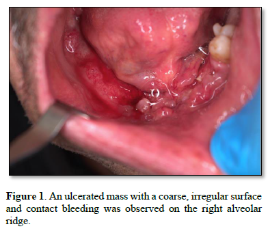
A 66-year-old male from San Fernando de Apure, with a history of tobacco use (DSM-5: 4; IPA: 5; Fagerström: 1), presented with a painful, bleeding tumor on the lower mandibular alveolar ridge (Figure. 1).

The patient reported progressive growth following a tooth extraction performed nine months earlier. He had received prolonged antibiotic therapy without improvement. Clinical findings included a 19-kg weight loss, anemia, and a history of gastritis secondary to chronic NSAID use. Occasional blood in the stool was reported.
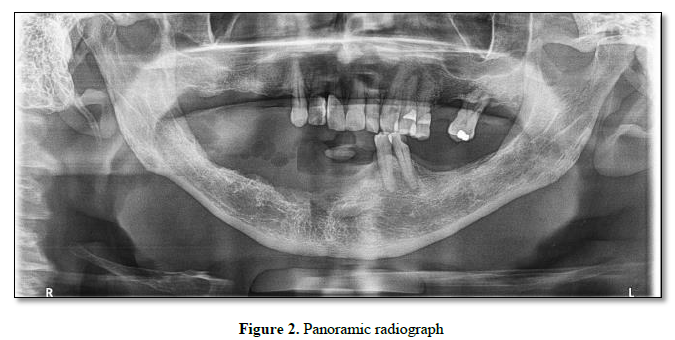
Radiographic studies included panoramic (Figure 2) radiography and cone-beam computed tomography. These revealed a radiolucent image corresponding to the post-extraction socket of tooth 43 and a radiopaque image showing mucosal thickening of the alveolar ridge in 4° quadrant, as well as increased trabeculation characteristic of the area.

Clinical examination showed an ulcerated exophytic lesion on the lower alveolar ridge. An incisional biopsy was performed. Imaging confirmed mandibular bone involvement. PET/CT did not reveal conclusive evidence of distant metastatic disease (Figure 3).

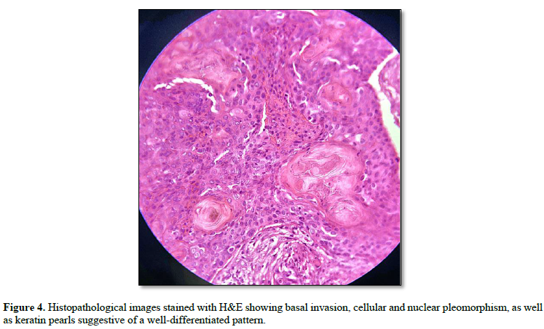
Histopathological analysis reported well-differentiated squamous cell carcinoma (Figure 4) with areas of dedifferentiation, basement membrane disruption, lamina propria invasion, angioinvasion, cellular and nuclear pleomorphism, abundant atypical mitoses, and keratin pearl formation. Immunohistochemistry for p16 was negative. The worst pattern of invasion was discohesive WPOI-3, with tumor heterogeneity and sparse inflammatory infiltrate in focal areas. Depth of invasion exceeded 2.5 mm. Invasion of adjacent osseous structures classified the lesion as T4a.

TREATMENT
Radiotherapy using VMAT (volumetric modulated arc therapy) was delivered to the lesion and cervical drainage areas. PTV: 7000 cGy at 200 cGy/day, for a total of 35 sessions (with immobilization mask).
Cisplatin 40 mg/m2 was administered concomitantly as a radiosensitizer for six cycles.

DISCUSSION
In resectable oral squamous cell carcinoma, surgery with adequate oncologic margins (including segmental or composite mandibulectomy when bone involvement is present), followed by adjuvant radiotherapy based on risk factors, is considered the standard approach to achieve locoregional control and survival (6).
However, in selected scenarios, definitive chemoradiotherapy or organ-preservation protocols (induction chemotherapy followed by chemoradiation) have been explored as alternatives to mutilating surgery, seeking a balance between tumor control and functional/aesthetic preservation (6).
In the present case, chemoradiotherapy was chosen instead of mandibulectomy, prompting discussion on selection criteria, available evidence, and the trade-off between oncologic outcomes and functional results.
The T4a classification was determined by mandibular bone invasion. Most historical series show that surgery followed by radiotherapy offers superior locoregional control rates in advanced oral disease, particularly when frank bone invasion is present (7).
Organ-preservation studies in the oral cavity have compared induction chemotherapy followed by chemoradiation with surgery plus selective radiotherapy, finding comparable overall and disease-free survival in carefully selected cohorts, although with higher risk of local failure in some subgroups (7).
Contemporary cohorts comparing definitive chemoradiotherapy with surgery plus radiotherapy in oropharyngeal carcinomas show that, when prognostic factors such as p16 status are adjusted, survival differences may diminish, reinforcing the importance of appropriate patient selection (8).
Recent comparative studies in oral cavity squamous cell carcinoma show that both surgery plus radiotherapy and definitive chemoradiotherapy improve baseline symptom burden, but toxicity profiles and functional sequelae differ: chemoradiotherapy may better preserve anatomy, though with risks of severe mucositis, xerostomia, and osteoradionecrosis (7).
Although this case exhibited histologic features associated with greater biological aggressiveness, the absence of detectable metastases allowed consideration of a functional-preservation strategy. In patients for whom resection would entail major mutilation, the organ-preservation literature in head and neck oncology supports considering non-surgical regimens, provided a reasonable probability of tumor control exists and surgical salvage is accepted in case of failure.
Current literature recognizes that various histopathological parameters-including worst pattern of invasion (WPOI), depth of invasion, and angioinvasion-provide complementary prognostic information. In this case, these factors were integrated into clinical decision-making (9).
CONCLUSIONS
This case highlights the importance of timely diagnosis of persistent lesions following dental procedures and the need for early histopathological evaluation. In selected patients, definitive chemoradiotherapy may constitute a therapeutic alternative aimed at preserving function and quality of life, always under strict clinical and radiological follow-up.
CONFLICT OF INTEREST
The author declares no conflict of interest related to the academic objectives of this publication.



No Files Found


Internationally Accepted
Share Your Publication :