
-
Publish Your Research/Review Articles in our High Quality Journal for just USD $99*+Taxes( *T&C Apply)
Offer Ends On
Publish Your Research/Review Articles in our High Quality Journal for just USD $99*+Taxes( *T&C Apply)
Offer Ends On
Juan Jose Soleri Cocco, M.J. Soleri Espada and Artigas Beltrán*
Corresponding Author: Artigas Beltrán, Professor of the Master’s Program in Implantology, University of Lleida, Spain.
Received: September 02, 2026 ; Revised: September 04, 2026 ; Accepted: September 06, 2026 ; Available Online: September 08, 2026
Citation: Soleri Cocco J J, M.J. Soleri Espada & Beltrán A. (2026) Transnasal Implants: Definition, Classification, Technique, And Use of New Monolithic Implants. J Oral Health Dent Res, 5(3): 1-20.
Copyrights: ©2026 Soleri Cocco J J, M.J. Soleri Espada & Beltrán A. This is an open-access article distributed under the terms of the Creative Commons Attribution License, which permits unrestricted use, distribution, and reproduction in any medium, provided the original author and source are credited.
Views & Citations
Likes & Shares
The rehabilitation of patients with atrophic maxillae has always been a challenge. Treatments involving large bone grafts, such as iliac crest grafts, and the use of zygomatic implants are not free from complications and adverse events. To prevent these problems and simplify treatment, several authors have proposed the use of tilted transnasal implants. This study consisted of a comprehensive literature review using a qualitative approach, with the purpose of explaining, obtaining in-depth knowledge of, and discussing transnasal implants. Different authors have described, rather than clearly defined, what is meant by “transnasal implants,” since different terminology is used for implants placed in the nasal region and both intranasal and extranasal approaches have been described. Camargo describes and classifies intranasal implants, whereas extranasal implants have not been classified; therefore, we propose the T.A.S.A. classification together with an integrated definition of transnasal implants anchored in the nasal region. Very few articles have been published on transnasal implants, most of them clinical case reports with short follow-up periods. Controlled studies with high methodological rigor are needed. All authors agree that the technique is predictable, that postoperative complications are transient, and that it should be included among the treatment options for atrophic maxillae.
Keywords: Transnasal implants; Frontonasal vestibulosinusal junction; Monolithic implants; Maxillary atrophy; Implant-supported fixed rehabilitation; Immediate loading.
RATIONALE
There are several reasons why we decided to select TRANSNASAL IMPLANTS: DEFINITION, CLASSIFICATION, TECHNIQUE, AND USE OF NEW MONOLITHIC IMPLANTS as the subject of study, among which the following are noteworthy.
Different authors agree that the technique is new and that, at present, very few articles have been published on transnasal implants, most of them clinical case reports with short follow-up periods, Gelpi F. et al. 2024 (1). In general, authors agree that the technique is predictable and an excellent alternative to zygomatic implants, either avoiding them or serving as an alternative to treatment with four zygomatic implants (quad zygoma), and as a complement to the placement of two implants. It may also be an alternative to large iliac crest bone grafts and Le Fort I techniques, both of which involve greater surgical risk, long recovery times, questionable medium- to long-term results, and higher economic costs.
With regard to juxtaosseous implants, also called subperiosteal meshes, this technique has been renewed compared with its historical form because of the use of the latest technologies in diagnosis, planning, use, and construction with new materials. However, there are no comparative studies regarding the success and suitability of one technique versus the other.
In atrophic posterior sectors with highly pneumatized sinuses, transnasal implants anchored to the anterior nasal wall can avoid sinus-lifting techniques with subantral grafts; combined with pterygoid implants, these areas can be rehabilitated.
We were also struck by the fact that publications on transnasal implants show a lack of information regarding the origin or “discoverer/first author,” a clear definition, and an integrated classification. Different authors have described rather than defined what is meant by “transnasal implants,” with important contributions from Jensen O.T. et al. 2010 (2,3,4), Camargo V.B. et al. 2019 (5,6), and Oh S. et al. 2023 (7).
We did not find an “author” of the technique, a clear definition, an integrated classification, a common area of bone anchorage, or a clear indication of which type of implant would be most suitable. Perhaps this situation warrants a consensus meeting to establish clear definitions.
This work aims to present a study showing that transnasal implants are a reconstructive method intended to restore the lost function of the stomatognathic system. However, the circumstances described above require a broader study and exhaustive analysis of the subject, relating the interactions among anatomy, radiographic diagnosis, anesthetic techniques, different surgical techniques, and their mutual influence. The aim is to provide clarity on the subject and establish guidelines regarding the definition, classification, diagnosis, and application of the different techniques; propose an appropriate anatomical area with minimal risk; and identify the implants most suitable for the technique, emphasizing in the final reflection the possibility of satisfactorily resolving patients’ clinical cases and contributing to their health and quality of life.
INTRODUCTION
The clinical demand for implant-based rehabilitation of tooth loss is highly relevant today. Tooth loss is accompanied by bone loss, resorption, and quantitative and qualitative atrophy that varies considerably according to different factors. Prolonged absence of teeth and conditions such as periodontal disease may produce severe resorption, leaving only a minimal amount of bone structure, J.J. Soleri Cocco et al. 2023 (8).
Bone loss in the maxilla may be partial or total. Grade IV–V–VI atrophy according to Cawood JI and Howell RA (1988) (9) represents the greatest rehabilitative therapeutic challenge. Resorption of the maxilla follows a centripetal and vertical pattern, Pietrokovski J. et al. (10), occurring much faster than in the mandible and producing an anterior and posterior crossbite situation.
Bone atrophy is accompanied by reduced bone quality, Lekholm U. Z.G.A. (1985) (11). The maxilla presents different bone qualities in its various areas. Although densities in the different regions are not “pure,” densitometrically the anterior region can be described as Type I–II, the premolar region as Type II–III, and the posterior molar region as Type III–IV. The latter generally presents low-density bone. These different densities condition the type, number, and ideal distribution of implants used for rehabilitation.
DIFFERENT OPTIONS AND ALTERNATIVES FOR MAXILLARY REHABILITATION
The different degrees of bone loss in partially or completely edentulous areas determine the implant-placement techniques and type of rehabilitation. When vertical (height) and horizontal (diameter) bone availability is adequate, basic surgical techniques for implant placement can be applied, with aesthetically and biomechanically ideal rehabilitation. When bone loss is greater, advanced surgical techniques are required, special implants with different types, morphologies, and sizes may be necessary, and more complex rehabilitation must be performed. This is a challenge requiring a high degree of experience from oral surgeons, implantologists, maxillofacial surgeons, and restorative dentists trained to rehabilitate these patients. Treatment options include:
TRANSNASAL IMPLANTS
Transnasal implants arose from the need to rehabilitate atrophic maxillae with insufficient bone that prevented the use of conventional endosseous implants, leading to the need for large bone grafts or the use of Quad Zygoma. As described by Bedrossian et al. in 2008 (20), radiographic areas of bone availability in the maxilla for dental implant placement included tilted conventional implants, zygomatic implants, or bone grafts. In the absence of bone volume in Zones 1, 2, and 3 of the maxilla, four zygomatic implants (Quad Zygoma) were indicated to rehabilitate patients with extreme bone atrophy as an alternative to grafting.
Currently, new implants such as compressive or basal monolithic implants and surgical techniques such as transnasal implants are available.
ANATOMICAL REVIEW
Maxillary bone atrophy forces us to place and anchor implants in areas distant from the residual alveolar ridges. The maxilla has areas called “buttresses,” Figun ME, Garino R. 2008 (21). These dense-bone buttresses form a protective framework around the multiple craniofacial cavities (orbit, nasal fossae, oral cavity, and paranasal sinuses), most of whose walls are fragile.
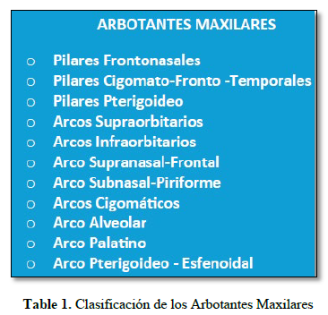
These buttresses, also called the SEIPEL TRAJECTORY SYSTEM, Seipel CM 1948 (22), distribute forces through the facial skeleton and have a strategic arrangement in each of the facial thirds, also called lines of force. These consist of cortical thickenings whose arrangement and trabecular condensation form reinforcement lines. According to their position in the bone, they are distinguished as pillars or columns when vertical and arches when horizontal, J.J. Soleri Cocco et al. 2022 (23).
Figure 1 / Table 1. Classification of maxillary buttresses.


The frontonasal pillar, the subnasal-piriform arches, and the basal alveolar arch are located on the basal maxillary bone and constitute the basic areas of bone anchorage for transnasal implants.
Important bony anatomical structures in this region in relation to transnasal implants include: residual alveolar bone; subnasal basal bone; the bony rim of the piriform notch; the nasal floor formed by the palatal processes of both maxillae joined at the midline; the anterior intermaxillary-vomerine bony crest; bilateral nasopalatine foramina forming the nasopalatine canal; and the bilateral frontal processes of the maxillae, which form the anterior wall of the nasal fossae on their internal surface. Approximately one third along this surface is the “bony bulla” of the anterior insertion of the inferior turbinate, with the opening of the lacrimal canal located posteriorly and superiorly. Its external surface is a continuation of the canine eminence and canine fossa, progressing superiorly to fuse medially with the nasal bones and superiorly with the frontal bone. Laterally it forms the internal suborbital rim and inferiorly forms part of the anterior surface, anterior rim, and part of the base of the truncated-pyramid-shaped maxillary sinuses. Figures 2–4.


TRANSNASAL IMPLANTS: WHERE ARE THEY PLACED OR ANCHORED?
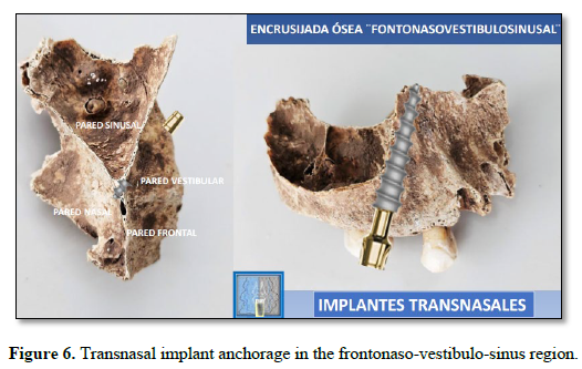
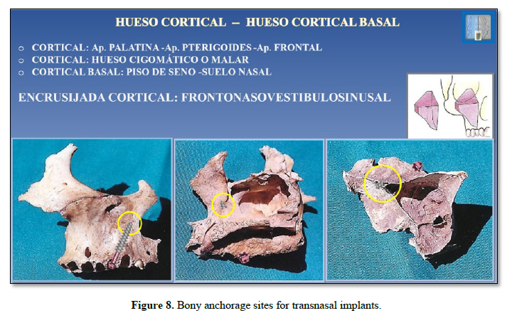
FRONTONASOVESTIBULOSINUSAL JUNCTION (Figures 5–8)
In the field of advanced dental implantology and maxillofacial surgery, we propose this area as a reference zone for strategic bone anchorage, fundamental for treating patients with severe maxillary atrophy without the need for massive bone grafts.
This compound term describes an anatomical bony area or “junction” in the middle third of the face. It indicates the convergence of several bony structures and air-containing cavities such as the sinus and nasal cavity:
Frontonasal process: process of the maxillary bone that ascends and joins the frontal bone and nasal bones.
Naso: nasal cavity.
Vestibule: external surface of the frontal process and part of the anterior wall of the maxillary sinus.
Sinusal: relating to the anterior border and part of the anterior surface of the maxillary sinus.
Frontonasalvestibulosinusal junction:
Anterior limit: anterior border of the frontal process and canine fossa.
Medial limit: external surface of the nasal fossa.
Posterior limit: anterior wall of the maxillary sinus.
Superior limit: anterior insertion of the inferior turbinate and opening of the lacrimal canal.
Inferior limit: canine fossa and canine apex.
In clinical practice, this region represents an anterior anatomical bony buttress. When a patient loses teeth and the alveolar ridge is resorbed, the bone in this area—especially the canine pillar and the floor/wall of the nasal fossae—remains relatively dense and stable. Surgeons take advantage of this remaining bone thickness to tilt or direct basal or compressive implants safely.
This is the clinical and methodological designation specifically given to the use of the frontonasalvestibulosinusal region for fixation of monolithic or single-stage implants, frequently basal or compressive implants. It is named according to biomechanical approaches and designs oriented toward strategic implantology. Instead of searching for vertical bone where it no longer exists, the proposed technique involves:
Bicortical anchorage: the implant crosses the remnant of the alveolar ridge and is firmly anchored in a second layer of hard cortical bone, such as the palatal process or floor of the nasal fossa.
Three-dimensional direction: implants are inserted in an inclined or divergent manner to avoid the maxillary sinus or take advantage of the nasogenian buttress without invading critical structures.
Immediate primary stability: fixation in these high-density bone areas allows the dentist to place a provisional or definitive fixed prosthesis immediately (immediate loading), restoring function and aesthetics to the patient within a few days.
In summary, the expression describes the surgical act of seeking support for and integrating implants into the dense anterior facial bony pillar (the junction of the nose, maxilla, and sinus), one of the strongest and most predictable areas after tooth loss.
This area permits placement both of the intranasal transnasal implants with medial/lateral inclination proposed by Camargo V.B. et al. 2019 (5) (Vanderlim Technique), and of the technique proposed by Jensen O.T. et al. 2010 (2), who described paranasally tilted implants with a lateral/medial inclination and called them transsinusal when anchored in the anterior border of the maxillary sinus over the frontal process, referred to as Point M, as well as another implant with medial/lateral inclination also anchored at Point M.


DEFINITION
Transnasal implants have been mentioned and described in different publications, with different techniques, anatomical sites, and positions, sharing the common feature of crossing the nasal floor and/or nasal walls to anchor one or more implants in basal bone with Type I density.
This raises the questions: What do we call transnasal implants? How can transnasal implants be defined?
Based on our 36 years of clinical and surgical experience in implantology and on the publications considered most important and key to the subject—Jensen O.T. et al. 2010 (2), Camargo V.B. et al. 2019 (Vanderlim Technique) (5), and Camargo V.T. et al. 2025 (6)—we propose the following integrated definition:
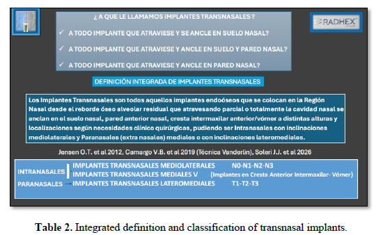
“Transnasal implants are all endosseous implants placed in the nasal region from the residual alveolar bony ridge that, by partially or completely traversing the nasal cavity, are anchored in the nasal floor, anterior nasal wall, anterior intermaxillary crest/vomer at different heights and locations according to clinical and surgical needs. They may be intranasal with mediolateral inclinations or paranasal (extranasal), medial or with lateromedial inclinations.” (Table 2).
INDICATIONS
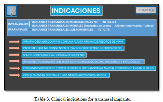
Transnasal Implants are indicated in patients with Grade IV–V–VI maxillary bone atrophy according to Cawood JI and Howell RA, 1988 (9); in All-on-Four or All-on-Six treatment plans using conventional implants; and in combination with pterygoid and zygomatic implants, following the distribution: Pterygoid Implant – Zygomatic Implant – Transnasal Implant – Transnasal Implant – Zygomatic Implant – Pterygoid Implant.
They avoid the use of four zygomatic implants (“Quad Zygoma”). They also avoid prostheses with posterior free ends (cantilevers).
They are indicated for patients who do not want bone grafting or for whom bone grafting is contraindicated. They are also indicated in patients with acute sinus pathology or recurrent chronic sinus pathology, poorly ventilated sinuses, and inadequate drainage (Table 3).
CLASSIFICATION
According to the integrated definition of transnasal implants, this is not a single procedure but several procedures involving a specific area within the nasal region. Several authors have used this anatomical area for implant anchorage but have called the procedures by different names, including angled implants, nasal implants, paranasal implants, and parasinusal implants. They describe the technique but do not provide a classification.
In mid-2016, Camargo described the Vanderlim Technique, registered the copyright and its scientific dissemination, Camargo V.B. et al. 2019 (Vanderlin Technique) (5). Since then, the Vanderlim transnasal implant technique has spread and been used by an increasing number of dentists worldwide.
To better guide procedures and reduce associated risks, this classification was developed for transnasal implant placement. It should be emphasized that this technique refers to the placement of intranasal implants and classifies them as follows. The NASA classification (acronym for Nasal Anatomical Systematic Approach) improves the surgical procedure. The surgeon must consider factors such as bone anatomy, the quality of the patient’s remaining bone, and the type of implant to be used. The exact location of the implant relative to the patient’s bony structures, alveolar crest height, shape of the nasal cavity, and availability of bone tissue for anchorage in the frontal process of the maxilla must be assessed to select the best technique.
NASA 0: the implant emerges at the crest in the region of the nasopalatine foramen.
NASA 1: the implant is in the central incisor region.
NASA 2: the implant is at the lateral incisor crest.
NASA 3: the implant is at the canine crest.
NASA 4: the implant is at the canine crest with exposed threads.
Jensen O.T. et al. 2010 (2) describe anchoring implants in the maxillary frontal-process area by angulating them, calling them parasinusal implants. They also describe implant placement in the anterior maxillary region with medial inclination, anchoring them in the anterior intermaxillary crest over the vomer, but these techniques were not classified.
Camargo V.B. et al. 2025 (6), based on Jensen, describe the placement of inclined lateromedial extranasal implants anchored at what they call five anchorage points in the paranasal transsinusal area (Figure 9).
 T.A.S.A. CLASSIFICATION
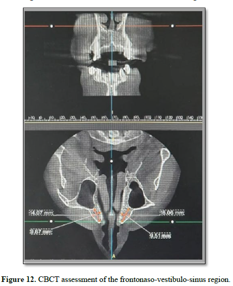
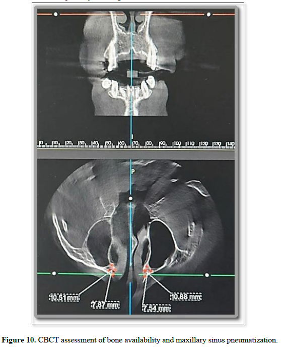
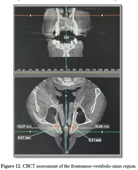
According to our experience and the literature reviewed, the placement of lateromedially angled implants starting from areas distant from 1.6–1.5–1.4 and/or 2.6–2.5–2.4 and anchored in the frontal process forming the anterior lateral wall of the nasal fossae has not been classified. Therefore, we propose the following classification of the different clinical and surgical possibilities according to the anatomy of the area, relating the shape and size of the maxillary sinus to the shape and size of the corresponding anatomical zone from the nasal floor to the frontal process and up to the anterior insertion of the inferior turbinate. This is the area described above and called the Frontonasalvestibulosinusal Junction (Figures 10–12). Bone availability is inversely proportional to the size and shape of the maxillary sinus (degree of sinus pneumatization), which conditions the implant-placement technique, implant type, and classification.
TRANSNASAL IMPLANTS (Extranasal), Lateromedial, T.A.S.A.
The T.A.S.A. classification (Transnasal Systematic Anatomical Utilization) is as follows:
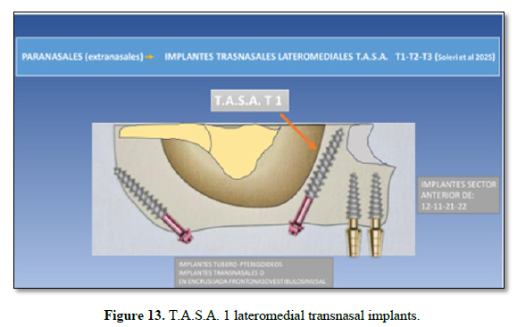
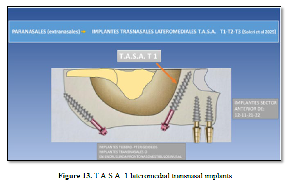
T.A.S.A. 1: (Small maxillary sinuses) The implant(s) travel lateromedially from the different zones or positions 1.6–1.5–1.4 / 2.4–2.5–2.6, crossing the remaining alveolar bone and reaching and anchoring in the Frontonasal vestibulosinusal Junction. The implant is completely surrounded by bone.
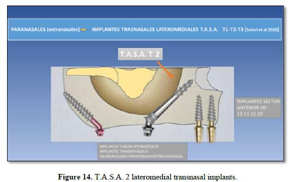
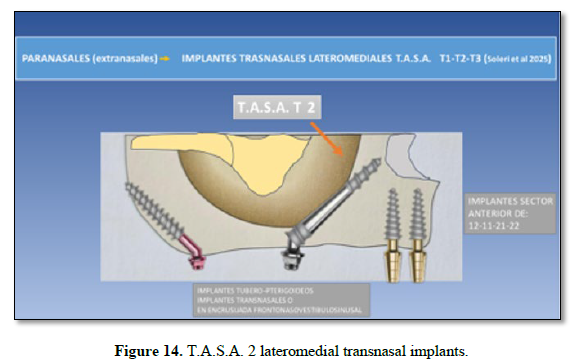
T.A.S.A. 2: (Medium-sized maxillary sinuses) The implant(s) travel lateromedially from the different zones or positions 1.6–1.5–1.4 / 2.4–2.5–2.6, partially crossing the remaining alveolar bone and contacting the anterior wall and anterior border of the sinus, reaching and anchoring in the Frontonasalvestibulosinusal Junction. The implant is partially surrounded by bone.
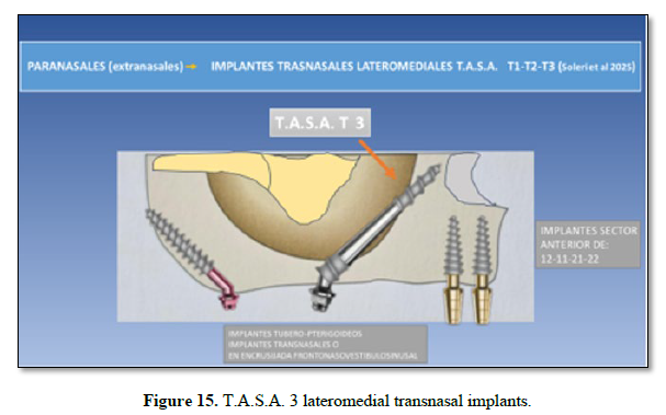
T.A.S.A. 3: (Highly pneumatized maxillary sinuses) The implant(s) travel lateromedially from the different zones or positions 1.6–1.5–1.4 / 2.4–2.5–2.6, partially crossing the remaining alveolar bone and entering the sinus, then penetrating again and anchoring in the Frontonasalvestibulosinusal Junction. The first third of the implant is surrounded by bone, the middle zone is “floating” intrasinusally, and the apical third penetrates and is anchored in bone. This situation may be combined with a sinus-lift technique using a rectangular transverse window made before implant insertion, followed by grafting to fill the cavity. (Figures 13–15).



SURGICAL TECHNIQUE
Surgery should be performed under aseptic conditions, with preparation of a sterile field in a dental office equipped for oral surgery.
Preoperative studies: A thorough preoperative assessment is necessary, including blood tests, complete blood count, coagulation profile, and liver profile; routine radiographic examination with panoramic X-ray, CT, and cone-beam computed tomography (CBCT) to determine the location, quality, and quantity in millimeters of available bone in the described anatomical area; a complete occlusal examination; evaluation of prosthetic space; and fabrication of radiographic and surgical positioning guides. These should determine the most favorable entry point in the maxilla and, consequently, the emergence point of the implant in the mouth, also evaluating its relationship with residual maxillary teeth and especially with their future mandibular antagonists.
It is also essential to assess the patient’s cooperation and mouth opening because the procedure is performed under locoregional anesthesia, and lack of cooperation and/or limited oral opening can seriously hinder or prevent the procedure.
Premedication: Antibiotic prophylaxis should be administered one hour before surgery, consisting of 2 g of amoxicillin or, in allergic patients, 600 mg of clindamycin + 500 mg of metronidazole. After surgery, it is recommended that all patients receive antibiotic treatment (amoxicillin 750 mg every 8 hours or clindamycin 300 mg every 8 hours + metronidazole 250 mg every 8 hours), an anti-inflammatory drug (ibuprofen 600 mg tablets every 8 hours), together with gastric protection (omeprazole 20 mg capsules every 24 hours).
Anesthesia: The surgical procedure is usually performed under locoregional anesthesia of the upper maxilla using 4% articaine (40 mg/mL) with epinephrine 1:100,000 (0.01 mg/mL), providing deep, rapid, and long-lasting anesthesia on both the maxillary vestibular and palatal surfaces. In complex cases or anxious patients, it is advisable for the anesthesiologist to additionally administer some form of intravenous sedation or even perform general anesthesia with nasotracheal intubation.
Incision: Using a No. 15 scalpel blade, an incision is made over the crestal mucosa of the posterior upper maxilla, accompanied by a distovestibular and distopalatal releasing incision at the first molar and extended to the corresponding contralateral area. A full-thickness mucoperiosteal flap is elevated with a periosteal elevator. Palatally, the palatal fibromucosa is elevated and the residual alveolar bone exposed. Vestibularly, the residual alveolar crest, subnasal area, piriform notch, anterior maxillary surface, and frontal process are exposed.
If intranasal mediolateral transnasal implants (Vanderlim Technique) are performed, Camargo V.B. et al. 2019 (5), the nasal fibromucosa must be elevated to expose the nasal floor and the internal anterior surface of the nasal fossae up to the anterior insertion of the inferior turbinate, called Point Z by Oh S. et al. 2023 (7).
If lateromedial extranasal transnasal implants are performed, either on the anterior vomerine intermaxillary crest, Jensen O.T. et al. 2010 (2), or on the Frontonasalvestibulosinusal Junction, Soleri J.J. et al. 2025 (24), intranasal elevation of the nasal fibromucosa is minimal and is performed only when necessary to verify the insertion of the implant apex.
In summary, intranasal mediolateral transnasal implants should be performed using open surgical techniques (with flaps), whereas extranasal lateromedial transnasal implants can be performed using either open or closed techniques (without flaps).
Implant-bed preparation: The steps for preparing the receiving bone bed of a transnasal implant are similar to those used in the standard technique for conventional two-piece endosseous implants: sequential drilling at an approximate medium-to-high speed of 1,000 rpm to create the cavity, followed by low-speed drilling and a torque of 50 Ncm to insert the implant, finishing with a manual wrench. The difference is the need for a kit of longer-than-usual instruments, including a handpiece or contra-angle with a straight head. To reach the basal cortical bone of the nasal floor and the frontal process of the maxilla, the bone bed must be drilled at the planned angulations.
Increased pressure on the handpiece or contra-angle is required because of the increased resistance of Type I basal cortical bone. Entry-and-exit drilling (“tapping technique”) and abundant cooling with chilled saline are recommended.
TRANSNASAL IMPLANTS (Extranasal), Lateromedial, T.A.S.A.: Based on our experience, the surgical technique for implant placement in this region may be open or closed (with or without a flap). With either option, implants may be placed freehand, using guided surgery, or using navigated surgery (Table 4).
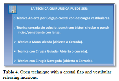
Table 4: Open technique with a crestal flap and vestibular releasing incisions; closed technique without a flap, using a circular punch or penetrating punch with a lance; freehand technique (open or closed); guided surgery (open or closed); navigated surgery (open or closed).
Bone drilling in this technique differs when monolithic implants are used. These significantly improve the technique because they are conical, have a soft compressive thread pitch ideal for Type I bone, can be bent to correct angulations up to 40°, and are suitable for immediate loading. Radhex Implants SLD-cm or SLD-c implants, with diameters of 3.5–4.5 mm and lengths of 17–25 mm, are implants that adapt particularly well to this technique (Figure 16).
The frequency of drilling is reduced, protecting poorly vascularized atrophic bone. Long drills or extension devices are used. The first drill is a 1.6-mm-diameter lanceolate drill, and the second is a 2.6-mm cylindrical drill for 3.5-mm implants; a third 3.6-mm drill is used when a 4.5-mm-diameter implant is selected. In many cases, depending on bone density, only progressively sized taps are used to expand and condense bone after the first and second drills.
Drilling in this technique has an approximate lateromedial inclination of 30–40° relative to the occlusal plane and another anteroposterior inclination of approximately 10–20°, depending on whether the starting position is 1.4–1.5–1.6 or 2.4–2.5–2.6, until the nasal floor and the Frontonasalvestibulosinusal Junction in the frontal process of the anterior lateral nasal wall are reached, where the implant apex is anchored.
Implant placement is initially performed with a contra-angle at 50 rpm and then continued with a manual ratchet wrench, or manually from the beginning using high torques of 40–50 Ncm. If the technique was open, the flap is repositioned and sutured. The previously fabricated immediate provisional fixed prosthesis is then placed, and occlusion is carefully checked. Figure 16.

OBJECTIVES
PRIMARY OBJECTIVE
To update the literature regarding patients with atrophic maxillae treated with transnasal implants and to evaluate this technique as an alternative.
SECONDARY OBJECTIVES
To evaluate quality of life and the long-term success of transnasal implants, and to determine the advantages, disadvantages, and risks that may be associated with performing this technique.
MATERIAL AND METHODS
This study consisted of a comprehensive literature review using a qualitative approach, with the purpose of explaining and obtaining in-depth knowledge about transnasal implants and discussing their definition, classification, clinical indications, contraindications, risks, effectiveness, feasibility, and survival rate.
We followed the methodology proposed by Whittemore and Knafl 2005 (25), consisting of: identification of the search problem; systematic searching of databases using previously defined inclusion and exclusion criteria; critical evaluation of selected studies; extraction, analysis, and interpretation of data; and synthesis of the findings in the literature.
As a reference framework, we used the PICO strategy and its variants. The PICO format represents successive components related to the Patient, Intervention, Comparison, and Outcome. These four components are fundamental elements of the research question and of constructing the question for an evidence-based literature search. The PICO question was used to derive and develop the title, objectives (primary and specific), keywords, and inclusion criteria.
P: Patients who have been treated with implants.
I: Transnasal implant-placement technique.
C: Patients treated with conventional implants or without implants.
O: To analyze the effectiveness, feasibility, and survival of transnasal implants.
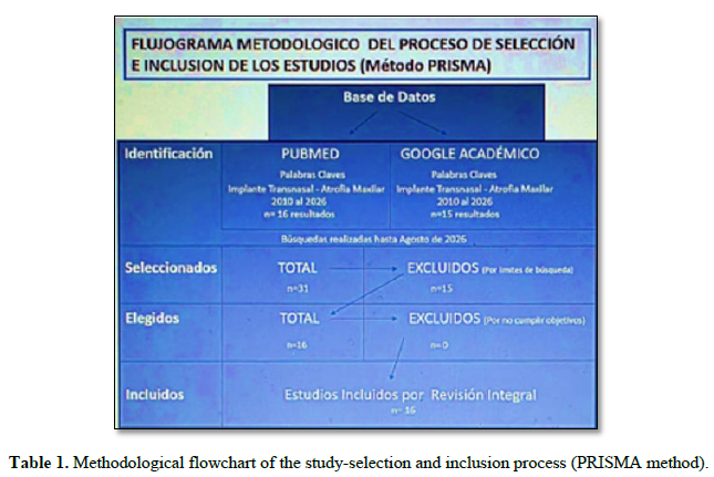
For the literature review, we focused the search on articles published in PubMed and Google Scholar between 2010 and 2026 (through August 2026). The search strategy used the following keywords: 1. Transnasal implants. 2. Maxillary atrophy.
Limits were established to make the search specific. For PubMed, the limits were: original articles of primary studies published in English or Spanish concerning the technique, classification, clinical cases, and case series; text availability: full text available (Free Full Text); species: humans.
The following were excluded: clinical trials; systematic reviews; meta-analyses; articles not freely available; and publications unrelated to the topic.
For Google Scholar, no limits were used. This database was used because it is very broad and also contains rigorous publications from different authors, institutions, languages, and countries that provide knowledge and evidence and may or may not be indexed.
After obtaining the results, duplicates were removed, and records were compared and excluded when, after reading the abstract, they were found not to be related to transnasal implants.
RESULTS
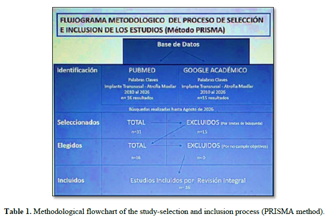
After studying all articles found for each keyword, a total of 31 results were obtained: 16 from PubMed and 15 from Google Scholar. After reading the abstract of each result, only 16 records were considered relevant for obtaining information: 13 from PubMed and 3 from Google Scholar. All 16 were used in the discussion. Tables 1 and 2 show the results obtained in this search.
DISCUSSION
Rehabilitation of patients with atrophic upper maxillae has always been a challenge for surgeons and restorative dentists. Treatments with large bone grafts, such as iliac crest grafts, and the use of zygomatic implants, especially in ZAGA P3 and P4 situations, Aparicio C. 2011 (37), as well as the Quad Zygoma configuration, are not free from complications and adverse events. These approaches may result in reduced health-related quality of life and may cause morbidity. To prevent these problems and simplify treatment, several authors have proposed the use of tilted paranasal (extranasal) implants, Jensen O.T. et al. 2010 (2), and transnasal (intranasal) implants, Camargo V.B. et al. 2019 (5).
Different authors have described rather than defined what is meant by “transnasal implants,” and different terminology is used for implants in the nasal region, including nasal, paranasal, transsinusal, and transnasal implants. Camargo V.B. et al. 2019 (5) calls implants placed intranasally transnasal implants, describes the technique (Vanderlim Technique), classifies it as N.A.S.A. 0–1–2–3–4, and patents it. However, in a 2025 publication, the same author, Camargo V.B. et al. (6), describes five anchorage points for transnasal implants; except for Point 1, which is for intranasal anchorage, Points 2–3–4–5 are for anchoring extranasal implants that are not related to the technique previously described.
Table 2. Characteristics of the articles, n=16.
|
No. |
Title |
Author/Year |
Database |
Journal |
|
1 |
The All-on-4 Shelf Maxilla. |
Jensen O.T. et al., 2010 (2) |
PubMed |
Journal of Oral and Maxillofacial Surgery |
|
2 |
Paranasal bone: the prime factor affecting the decision to use transsinus vs zygomatic implants for biomechanical support for immediate function in maxillary dental implant reconstruction. |
Jensen O.T. et al., 2014 (4) |
PubMed |
Int J Oral Maxillofac Implants |
|
3 |
Extra-Long Nasal Wall-Directed Dental Implants for Maxillary Complete Arch Immediate Function: A Pilot Study. |
Nicoli G. et al., 2019 (26) |
PubMed |
Oral Maxillofac Surg Clin North Am |
|
4 |
Transnasal Implant (Vanderlim Technique) as an option to the second zygomatic implant. |
Camargo et al., 2019 (5) |
Google Scholar |
Coppedê A. (ed.). Clinical solutions for total implant-supported rehabilitations without bone grafts. São Paulo: Quintessence, 2019. |
|
5 |
Consensus Regarding 16 Recognized and Clinically Proven Methods and Sub-Methods for Placing Corticobasal® Oral Implants. |
Antonina Idhe et al., 2020 (27) |
PubMed |
Ann Maxillofac Surg. |
|
6 |
Extra long transnasal implants as alternative for Quad Zygoma: case report. |
Almeida P.H. et al., 2021 (28) |
PubMed |
Annals of Medicine and Surgery |
|
7 |
Trans-sinus dental implants, for immediate placement when insufficient alveolar height is present: an alternative to zygomatic implants – surgical case series. |
Aalam A.A. et al., 2023 (29) |
PubMed |
Annals of Medicine and Surgery |
|
8 |
Case report: utilization of Z-Point fixture “Trans-nasal” implants. |
Oh S. et al., 2023 (7) |
PubMed |
Ann Med Surg (Lond) |
|
9 |
Treatment of Severely Atrophic Maxilla by Using Zygomatic, Pterygoid, and Transnasal Implants. |
Şahin O., 2024 (30) |
PubMed |
J Craniofac Surg. |
|
10 |
All-on-4 Hybrid with Extra-Long Transnasal Implants: Descriptions of the Technique and Short-Term Outcomes in Three Cases. |
Nunes M.N. et al., 2024 (31) |
PubMed |
Journal of Clinical Medicine |
|
11 |
The use of the Transnasal implant associated with the zygomatic implants in the treatment of atrophic maxilla: A Case Report. |
Sales P. et al., 2024 (32) |
Google Scholar |
The European Journal of Stomatology, Oral and Facial Surgery |
|
12 |
Trans-nasal dental implants: indication and the report of 10 cases. |
Eshoiee N. et al., 2025 (33) |
PubMed |
Annals of Medicine and Surgery |
|
13 |
A retrospective multicentric study of 52 nasal and transnasal implants in 31 severely atrophic patients to reduce anterior cantilever bending in full arch implant-supported fixed rehabilitations. |
Gelpi F. et al., 2025 (1) |
PubMed |
International Journal of Oral & Maxillofacial Implants |
|
14 |
Feasibility of transnasal dental implant placement determined according to PLACATE guidelines. |
Holtzclaw D. et al., 2025 (34) |
Google Scholar |
Dental Research and Oral Health |
|
15 |
An Innovative Technique to Immediately Load a Severely Atrophic Maxilla Without Zygomatic Dental Implants Using the 3B-TB Protocol. |
Chandrasekaran M. et al., 2025 (35) |
PubMed |
Cureus. 10.7759/cureus.93170. |
|
16 |
Transnasal Implants as an Alternative to Quadzygoma in the Rehabilitation of Extremely Atrophic Maxillae: Long-Term Results of the Vanderlim Technique Cases Report. |
Camargo V.B. et al., 2026 (36) |
PubMed |
Int J Oral Maxillofac Implants. |
Although the term “transnasal implants” and the classification can be attributed to Camargo V.B. when the implants are intranasal and medially/laterally inclined, he now accepts that they can also be extranasal, lateromedially inclined, and anchored at different points in the paranasal region.
Of the 16 publications included in this review dealing with transnasal implants, 11 concern paranasal extranasal lateromedial implants and 5 concern intranasal mediolateral implants: Almeida P.H. et al. 2021 (28), Oh S. et al. 2023 (7), Holtzclaw D. et al. 2025 (34), and Camargo V.B. et al. 2019 and 2026 (5,36).
After reviewing the 16 publications included in this review, no author had classified the full range of these implants, nor did we find a clear and integrated definition of transnasal implants. Therefore, as explained in this article, we propose an integrated definition encompassing the different possibilities for implant placement in the nasal area: “Transnasal implants are all endosseous implants placed in the nasal region from the residual alveolar bony ridge that, by partially or completely traversing the nasal cavity, are anchored in the nasal floor, anterior nasal wall, anterior intermaxillary crest/vomer at different heights and locations according to clinical and surgical needs. They may be intranasal with mediolateral inclinations or paranasal (extranasal), medial or with lateromedial inclinations.”
Because there is no classification for the placement of extrasinus transnasal implants with lateromedial inclination, as explained above, we propose the classification: TRANSNASAL IMPLANTS (Extranasal), Lateromedial, T.A.S.A. (Transnasal Systematic Anatomical Utilization): T.A.S.A. 1–2–3.
Many authors agree that the technique is relatively new and that the number of publications on transnasal implants is currently very small. Most are clinical case reports with short follow-up periods, including one clinical case each by Almeida P.H. et al. 2021 (28), Oh S. et al. 2023 (7), and Sales P. et al. 2024 (32). Aalam A.A. et al. 2023 (29) is notable because the title describes a clinical case series, but the article itself reports only two cases. Nunes M.N. et al. 2024 (31) reports three clinical cases, Eshoiee N. et al. 2025 (33) reports ten clinical cases, and Gelpi F. et al. 2025 (1), in a retrospective study, reports placement of 52 transnasal implants in 31 patients with 100% success. Camargo V.B. et al. 2026 (36) presents the largest published series to date, including clinical data from 226 transnasal implants placed in 121 patients treated at five different clinical centers worldwide, with follow-up ranging from 1 to 8 years. No early or late failures were observed. All implants met the survival criteria.
In this study, the survival rate of transnasal implants was 100%.
Regarding diagnosis and planning, there is consensus that digital 3D volumetric tomography (CBCT) with virtual planning software should always be used, allowing surgical guides to be fabricated or navigated surgery to be performed. CBCT is essential, Holtzclaw D. et al. 2025 (34), to study the remaining bone and anatomical structures individually because there is substantial variability between patients and even between the right and left sides of the same patient.
Regarding indications, there is broad consensus that transnasal implants avoid bone grafting. With respect to rehabilitation planning, some authors advocate their use to avoid zygomatic implants, arranging them in an M configuration with four implants or a Double-M configuration with six implants, as described by Jensen O.T. et al. 2010 and 2014 (2–4), Nicoli G. et al. 2019 (26), Aalam A.A. et al. 2023 (29), Nunes M.N. et al. 2024 (31), and Chandrasekaran M. et al. 2025 (35). Evaluating transnasal implants as an alternative to zygomatic implants is very important when planning rehabilitation, although medium- to long-term comparative studies are lacking, as is also the case with new-generation juxtaosseous implants.
Other authors use them to avoid Quad Zygoma, complementing two zygomatic implants. They therefore perform six-implant rehabilitations consisting of 2 transnasal, 2 zygomatic, and 2 pterygoid implants, as reported by Camargo V.B. et al. 2019 and 2026 (5,36), Antonine Idhe et al. 2020 (27), Almeida P.H. et al. 2021 (28), Şahin O. 2024 (30), Sales P. et al. 2024 (32), and Eshoiee N. et al. 2025 (33). It has also been described that transnasal implants avoid distal free-end prostheses (cantilevers), Gelpi F. et al. 2025 (1).
All authors agree on immediate-loading prostheses over transnasal implants. Authors such as Gelpi F. et al. 2025 (1) delay immediate loading when insertion torque is below 50 Ncm, whereas Camargo V.B. et al. 2026 (36) performs immediate loading with torque from 35 Ncm.
They are indicated not only in completely edentulous patients with maxillary bone atrophy but also in partially edentulous patients. When there is a distal free end, the combination of transnasal and pterygoid implants avoids and bypasses the maxillary sinus and is a valid option for highly pneumatized sinuses, sinuses with chronic or acute pathology, poorly ventilated sinuses, and patients who do not accept sinus lifts or bone grafts.
Contraindications include the general contraindications inherent to any type of implant, patients with sufficient bone for conventional implants, and lack of operator training in these techniques. Nevertheless, it should be emphasized that this technique simplifies treatment of patients with atrophic maxillae and allows a greater number of surgeons to perform it in private practice under local anesthesia.
Disadvantages and risks: the intranasal transnasal implant technique with mediolateral inclination (Vanderlim Technique), Camargo V.B. et al. 2019 (5), which advocates anchoring the implant apex in the bulla of the anterior insertion of the inferior turbinate called Point Z by Oh S. et al. 2023 (7), has the disadvantage that the opening of the lacrimal canal is immediately and very close posteriorly and superiorly in the middle meatus, at approximately 3 mm, Holtzclaw D. et al. 2025 (34). It may therefore be invaded and injured, causing perforation of the nasolacrimal duct and serious consequences such as dacryostenosis, which can in turn cause epiphora and dacryocystitis.
Therefore, meticulous preoperative anatomical assessment and planning are necessary to ensure the safe use of transnasal dental implants. Evaluation of the specific bony structures with which the implants interact is also required to ensure appropriate case selection and implant survival.
This methodology also involves elevation of the nasal fibromucosa, which may tear during the procedure, exposing the implant and the biomaterials used as fillers. Implants placed with this technique and classified as N.A.S.A. 0–1–2 literally cross the lumen of the nasal opening and may produce transient paresthesia, tingling, and a “needle-like” sensation in the nasal ala; postoperative respiratory difficulty in patients with a deviated septum; and fistula due to excess.
The extranasal transnasal implant technique with lateromedial inclination is simpler, and the implant apex is anchored lower in the Frontonasalvestibulosinusal Junction, avoiding all of the risks described above, Jensen O.T. et al. 2010 and 2014 (2–4), Chandrasekaran M. et al. 2025 (35), Soleri J.J. et al. 2025 (24). Nevertheless, either technique may, for different reasons, result in accidental perforation of anatomical structures, secondary or recurrent sinusitis, exposure and premature mobility of the implant, and peri-implantitis.
Regarding the type of implant used for these transnasal implant techniques, almost all included authors describe conventional two-piece cylindrical endosseous implants with lengths ranging from 16 to 25 mm and diameters of approximately 3.5–4.5 mm, in which the apex has the same diameter as the body or is slightly smaller.
We agree with Antonine Idhe et al. 2020 (27) and Chandrasekaran M. et al. 2025 (35) on the use of monolithic implants. Antonine I. describes the use of Idhe® monolithic implants and Chandrasekaran M. the use of Bioline® monolithic implants, whereas our experience concerns Radhex Implant® monolithic implants. These implants have a design suitable for anchorage in basal bone, with a self-tapping thread, a conical shape, and, although the diameters and lengths used are similar, they terminate in a narrow apex of 1–1.5 mm, which is ideal for reaching anatomically compromised areas with lower risk.
All authors agree that the technique is predictable and an excellent treatment option, with success rates between 95% and 100% and satisfactory longevity. Sales PH. 2025 (38) describes an effectiveness of 97.2%. No statistically significant difference in effectiveness was found when transnasal implants were compared with conventional implants (p = 0.14, I² = 0%), nor was a statistically significant difference found in marginal bone loss between the groups (p = 0.47, I² = 91%). None of the studies analyzed presented a low risk of bias, considering that this is a relatively new technique with few comparative studies, short series, and short- to medium-term follow-up.
CONCLUSIONS
The use of transnasal implants in the rehabilitation of edentulous maxillae represents an appropriate option with a high success rate and should be included among the treatment options for atrophic maxillae.
There is a reduced surgical risk compared with other techniques, and the rehabilitation treatment is simplified.
The technique can be performed by a greater number of dentists in the dental office under locoregional anesthesia.
Transnasal implants are not a technique; they are essentially a concept for implant placement in the nasal region involving three well-defined techniques: intranasal transnasal implants with mediolateral inclination; infranasal medial/vomerine transnasal implants; and extranasal lateromedial transnasal implants (paranasal).
The use of monolithic implants substantially improves these techniques compared with conventional two-piece cylindrical implants.
However, the small number of controlled studies on the subject and the high risk of bias are important factors that must be considered before choosing this therapy.
New controlled studies with high methodological rigor are necessary to determine the effectiveness of transnasal implants in the rehabilitation of atrophic maxillae.
DECLARATION OF CONFLICTS OF INTEREST
The author declares that there is no conflict of interest.








No Files Found


Internationally Accepted
Share Your Publication :