
-
Publish Your Research/Review Articles in our High Quality Journal for just USD $99*+Taxes( *T&C Apply)
Offer Ends On
Publish Your Research/Review Articles in our High Quality Journal for just USD $99*+Taxes( *T&C Apply)
Offer Ends On
Philip L Millstein DMD, MS*
Corresponding Author: Philip L Millstein, Department of Restorative Dentistry, Harvard School of Dental Medicine, Boston, Massachusetts, USA.
Received: September 01, 2026 ; Revised: September 04, 2026 ; Accepted: September 06, 2026 ; Available Online: September 07, 2026
Citation: Millstein PL. (2026) Occlusal Contact Marking Indicators. They All Differ. J Oral Health Dent Res, 5(3): 1-3.
Copyrights: ©2026 Millstein PL. This is an open-access article distributed under the terms of the Creative Commons Attribution License, which permits unrestricted use, distribution, and reproduction in any medium, provided the original author and source are credited.
Views & Citations
Likes & Shares
Occlusal contact indicators differ in marking quality. Oral health and dental stability are often judged by viewing intraoral contact markings. The quality of contact reproduction is unknown.
Keywords: Occlusion, Occlusal contact, Marking indicators, Electronic systems.
Marking indicators mark upon contact of opposing teeth. They are disposable. Dentists are taught to use them once or twice and then to discard them. The cost for each indicator may appear minimal but when multiplied two or three hundred thousand times a day the profit becomes significant. Students in dental school are shown what indicators to use and how to use them. As practitioners they may be introduced to new products. Whether new products are better than older ones is unknown. There is no definitive way to make an educated choice of what indicator to use because there are no testing standards. How does a clinician decide what indicator to choose? A suggestion by a colleague is usually sufficient. A tooth marking is a by product of an indicator Figure 1.

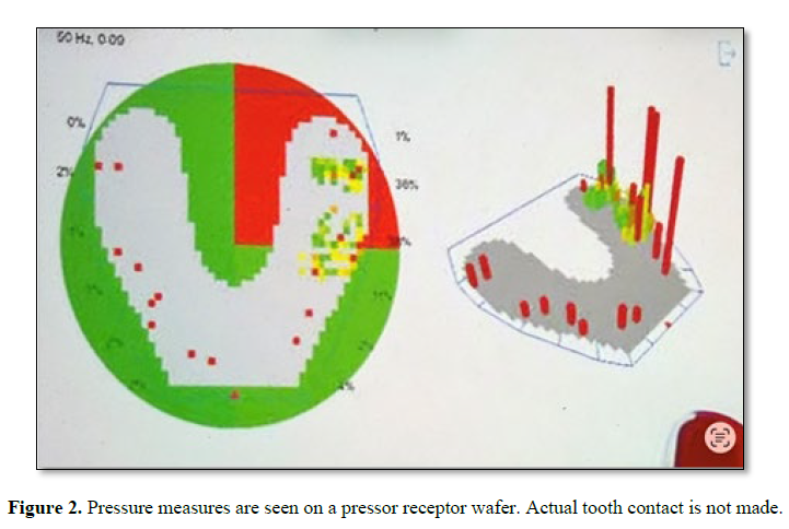
There are many marking systems (1). Electronic systems are available that record pressure points along with contact timing in pressure sensitive bite wafers Figure 2. The results are converted into estimated contact areas (2). Actual tooth contact is not made. Some occlusal treatments are built on using the pressure sensitive wafers (3). Fast disclusion is advocated as a sign of occlusal health (4). The faster the disclusion time the more functional the occlusion. Cuspal obstruction is minimized. Removal of tooth surfaces using the wafer as a disclusion guide may also eliminate occlusal contact stops which prevent “bite” collapse (5).
The long term results are unknown. Tooth to tooth contact can only be recorded using contact indicators. Contact indicating strips vary in color, thickness, and flexibility. Some are smudge proof. Indicators mark but the markings vary since there are so many different kinds of indicators (6). A clinician must use an indicator. Choice of a marking system is subjective. Occlusal stability depends upon occlusal contact (7).
Occlusion may be the most significant factor in sustaining dental health (8).
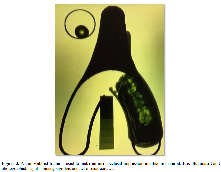
The only way to determine the veracity of a marking system is to use it. Personal evaluation of a marking system requires making an inter occlusal record and analyzing it (9). A silicone impression material is placed into a triple tray (Premier Dental) and a patient bites hard Figure 3.


Upon set the impression is removed and placed onto an illuminated light box and photographed. The record is permanent. Viewing the photo will provide some idea of indicator efficiency (10). Further analysis using image analysis will provide a complete picture of a true occlusal contact and the surrounding area Figure 4.

This is a true reproduction of an occlusal contact. The size and shape of the red contact area has not been distorted by the impression. Comparisons of various markings can be made using the impressed contact as a guide. An occlusal adjustment whether minimal or maximal requires planning. This includes using appropriate cutting instruments. Contact markings dictate tooth reduction. Large marks require gross reduction; small marks require minimal reduction. The questions remain. Are the marks precisely located and is the size of contact true or is it determined by the indicator? Gross tooth reduction without planning leads to occlusal instability. Unplanned occlusal adjustment is performed daily. Bite collapse is common. Prevention other than bite guard intervention is rare.



No Files Found


Internationally Accepted
Share Your Publication :