
-
Publish Your Research/Review Articles in our High Quality Journal for just USD $99*+Taxes( *T&C Apply)
Offer Ends On
Publish Your Research/Review Articles in our High Quality Journal for just USD $99*+Taxes( *T&C Apply)
Offer Ends On
Marwan Abou-Rass*, DDS, MDS, Ph.D.
Corresponding Author: Marwan Abou-Rass, USC. Prof Emeritus, Founder of AbouRass Endodontics Academy (www.abourass.com)
Received: July 28, 2026 ; Revised: July 29, 2026 ; Accepted: July 31, 2026 ; Available Online: August 03, 2026
Citation: Abou-Rass M. (2026) The 4R Operational Diagnosis Protocol: Part 4: R4 – Restorative and Tooth Structure Assessment. J Oral Health Dent Res, 5(3): 1-17.
Copyrights: ©2026 Abou-Rass M. This is an open-access article distributed under the terms of the Creative Commons Attribution License, which permits unrestricted use, distribution, and reproduction in any medium, provided the original author and source are credited.
Views & Citations
Likes & Shares
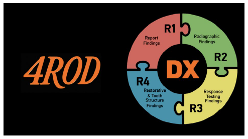
Introduction: Abou-Rass presents the final component of the 4R Operational Diagnosis (4ROD) Protocol: Part 4 (R4), a structured phase that integrates restorative and tooth structure assessments during coronal cleanout to reach a definitive diagnosis.
Methods: R4 is executed under rubber dam isolation using a dual framework:
Result:
Conclusion: The R4 phase operationalizes the 4ROD protocol by eliminating restorative and structural ambiguities prior to treatment, ensuring long-term endodontic success.
Keywords: 4ROD Protocol | Restorative Assessment | 2023 FDI Criteria | Rule-Out Tooth Crack (ROTC) | EROIC Framework | Tooth Structure Cracks (TSCs) | 10 ADIs-RIS | Pulp and Perio Response Testing | Coronal Cleanout | Non-Carious Cervical Lesions (NCCLs)
INTRODUCTION
The 4R Operational Diagnosis Protocol is an operational, structured, sequential, biologically based, evidence-based diagnostic system developed by AbouRass 1982 that evaluates the tooth, pulp-Dentin complex, and periodontium through the patient’s Report (R1), Radiographs (R2), Response to testing (R3), and restorative and tooth structure status (R4) to reach a definitive operational diagnosis and treatment plan.
R4 serves as the key phase for making definitive diagnoses, combining findings from R1, R2, and R3. It helps develop a problem list and a treatment plan and prepares the case for presentation to the patient.
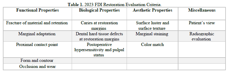
R4 includes two parts: the restorative assessment, based on the 2023 FDI criteria for restoration evaluation, and the tooth structure assessment, which addresses the main pathological conditions relevant to endodontics from a biological and clinical perspective.
The articles R1: Patient Report and Rapport, R2 – The 10 ADIs Radiographic Interpretation System (10 ADIs-RIS), and R3 Mastering R3 – Pulp and Perio Response Testing can be reviewed at the following links:
R1: https://www.genesispub.org/resource/images/articles/pdf755.pdf
R2: https://manuscriptscientific.com/images/2ddd2976aac44b31e58c5744aa8def17.pdf
R3: https://manuscriptscientific.com/images/17fd69979925a4b1b5ff518347cade84.pdf
Q1. What is the Clinical Importance of the R4 Phase in the 4ROD Protocol?
Operationalization represents the core power of the 4ROD protocol. This power is most evident in the R4 phase, following the clinical mantra: let’s open and see. Here, the rubber dam isolation and aseptic field are set up in preparation for a thorough coronal cleanout.
R4 marks the critical phase where existing restorations are carefully removed to expose hidden cracks on the cavity floor, cusps, foundations, and line angles. During this process, crowns on ETT are removed to detect recurrent decay, microleakage stains, pulp chamber perforations, and untreated canal openings.
In R4, a new periapical or bitewing radiograph is taken, and the findings from R1, R2, and R3 are reviewed, with the coronal clean-out results considered. A final diagnosis is then made, leading to the creation of a problem list and a treatment plan.
The R4 stage includes two parts: The Restorative Assessment component of R4 adheres to the 2023 standards set by the FDI World Dental Federation, regarded as the standard for evaluating both direct and indirect restorations and The Tooth Structure Assessment component of R4 examines key acquired conditions affecting tooth structure, pulp, periapical, and periodontal health, including tooth structure cracks, discoloration, surface wear, and dental anomalies.
Q2. Should Endodontic Treatment Be Performed Through Existing Restorations Regardless of Their Quality?
Proceeding with treatment through an existing crown or restoration without first verifying its integrity may lead to overlooking the actual cause of the problem. It could transform a technically correct endodontic procedure into a long-term failure.
Performing endodontic treatment through crowns and existing restorations is typically justified by financial constraints, convenience, or improved rubber dam isolation. Still, these reasons are unacceptable from biological, clinical, and practice-management perspectives. Concerns about patient cost do not warrant overlooking hidden disease, and convenience should never outweigh the importance of accurate diagnosis, patient safety, and responsible treatment planning.
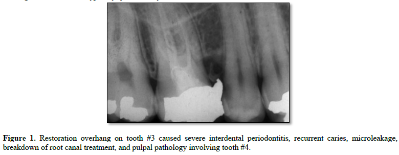
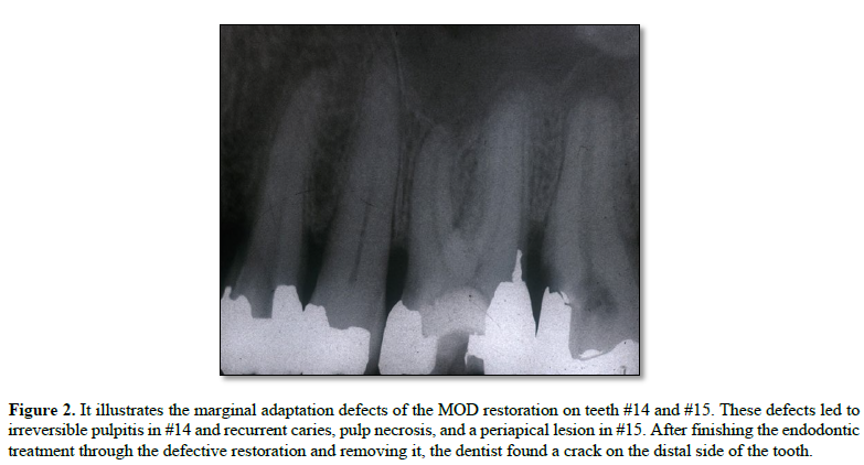
In my early clinical practice, I followed traditional methods, only to often encounter regret and surprise. I regret causing perforations, missing canal orifices hidden beneath crown margins, failing to diagnose cracks, and recurrent caries after crown removal. Sometimes the damage was so extensive that the tooth could no longer be repaired.
The answer to your question is no! That's why I made restoration assessment a key part of the tooth structure evaluation in R4.
4ROD Clinical Guideline:
Endodontic treatment must not be performed through existing intracoronal restorations or crowns that fail to meet FDI functional, biological, and aesthetic criteria.
Q3. What Criteria Do You Employ to Assess Existing Restorations in Teeth That Need Endodontic Treatment or Retreatment?
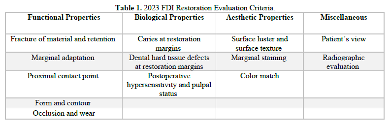
Currently, the 2023 FDI criteria serve as the standard for assessing both direct and indirect restorations. Developed in 2010 by a team of international educators and researchers in restorative dentistry and dental materials, these criteria have been refined over 13 years of global testing. While the 2010 criteria emphasized aesthetics first, followed by biological and functional aspects, the 2023 criteria reorganized this sequence. Additionally, while the 2010 version focused on criteria, the 2023 update replaced criteria with properties.
FDI-Functional Properties
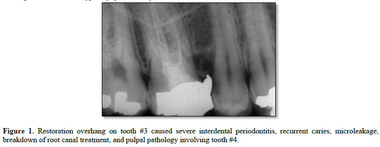
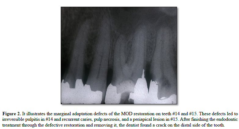
Marginal adaptation is essential for assessing the success of restorations and crowns. A well-fitted margin ensures a seamless, smooth transition between the restoration and the tooth. Deficiencies in marginal adaptations, such as open margins, overhangs, and horizontal and vertical discrepancies, can lead to recurrent caries, gingival inflammation, crown cementation failure, or problems with the restorative material. Additionally, the functional criteria encompass restoration shape, contour, and proximal contact points, as flaws in these areas can cause biological and aesthetic issues, as well as pulpal or endodontic complications.
FDI-Biological Properties
CAR (Caries at Restoration margins) indicates secondary or recurrent caries caused by poor marginal fit and other deficiencies. This biological failure directly damages tooth structure. Often small and asymptomatic, these lesions are hidden and only visible through radiographs in advanced stages. While usually without symptoms, CAR can cause dentinal hypersensitivity and early pulpitis as it worsens.
In endodontically treated teeth, bacterial microleakage associated with caries leaks in the pulp chamber can compromise the root canal filling and promote biofilm formation, accelerating the failure of endodontic treatment.
In addition to these endodontic problems, advanced CAR also causes interdental gingivitis and periodontitis defects that are challenging to detect radiographically.
FDI-Aesthetic Properties
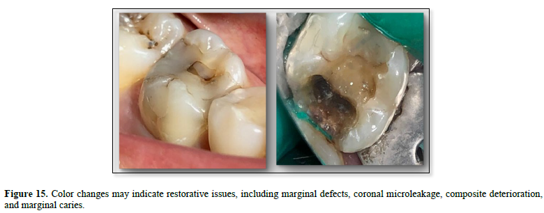
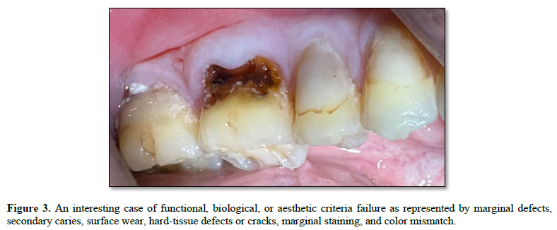
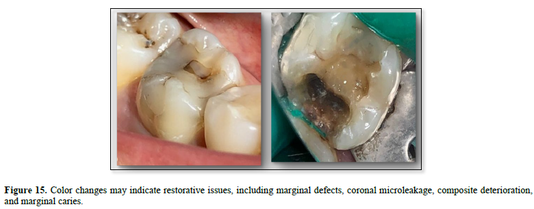
Meeting the aesthetic criteria in a restoration means that both the Biological and Functional Criteria are satisfied. This is because marginal staining and color changes are primarily caused by coronal microleakage, which arises from underlying issues such as Insufficient marginal adaptation, deterioration of restorative materials, ongoing endodontic treatment failure, and root resorption problems.
Meeting the aesthetic criteria in a restoration means that both the Biological and Functional Criteria are satisfied. This is because marginal staining and color changes are primarily caused by coronal microleakage, which arises from underlying issues such as Insufficient marginal adaptation, deterioration of restorative materials, ongoing endodontic treatment failure, and root resorption problems.
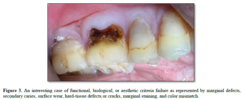
Q4. How Do You Compare the Evaluation Process for Intracoronal Restorations with That for Crown Restorations Before Endodontic Treatment?
Removing intracoronal restorations during the coronal clean-out is a required clinical guideline to uncover underlying decalcified dentin, concealed cracks, recurrent caries, cusp foundation cracks, and to expose the tooth structure for comprehensive assessment.
Crowns require a more careful evaluation. They should be assessed according to the current FDI criteria; if they are considered clinically acceptable, root canal treatment can proceed through them. To avoid complications, I recommend referring the patient to an endodontist with specific instructions to maintain the crown.
If the crown is of poor quality or does not meet the evaluation criteria, it should be removed before treatment. This helps accurately identify the true cause of pulp disease or endodontic failure, thereby simplifying the endodontic procedures.
In my experience, when assessing the quality of older crown restorations associated with pulp issues or unsuccessful endodontic procedures, only a small number are worth keeping, as the crown itself often contributed to or caused the problem we're addressing.
Performing endodontic retreatment through crowns with an existing access opening involves considerable risks. Despite being a common practice, I personally avoid this method.
4ROD Clinical Guideline:
Intracoronal restorations must be completely removed during coronal cleanout to expose tooth structure and allow accurate restorative and endodontic assessment, diagnosis, and treatment planning.
Q5. What Common Restoration Deficiencies are Encountered in Practice, and How are They Scored?
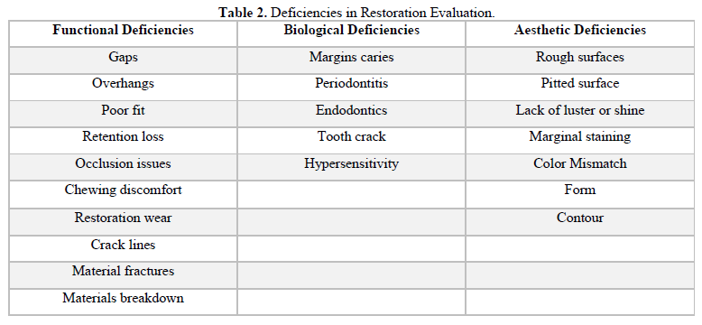
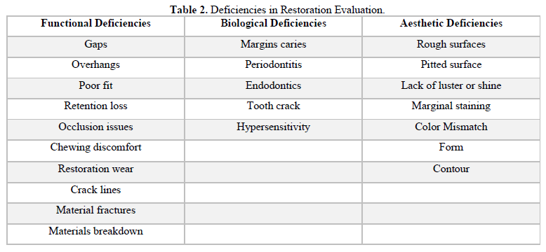
Table 2 outlines potential deficiencies commonly encountered in direct and indirect restorations. Endodontically speaking, the emphasis is on crown restorations. The assessment results determine whether to retain or remove the crown restoration, a key decision in endodontic treatment planning.
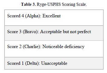
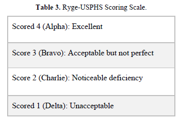
Previously, we used Ryge's criteria from the 1980s and the modified USPHS guidelines from the 1990s. (Table 3) These early standards had limitations because they did not include patient feedback on function, symptoms, or tissue responses. They also lacked emphasis on radiographic evaluation of restorations.
Relying solely on clinical assessment results in a loss of about 50% of diagnostic information. Even the 2010 FDI five-point scale—from excellent to poor—was less definitive than the 2023 criteria we now employ. Today, the restoration assessment is conducted as a separate procedure, following this process.
Restoration Assessment Requirements and Instruments:
In the 4ROD protocol, I use the current 2023 World Dental Federation (FDI) criteria, along with the scoring scale listed in the 4ROD Clinical Guideline.
4ROD Clinical Guideline: Restoration Assessment
R1: Record patient feedback on restoration function and symptoms.
R2: Bitewing radiograph to evaluate marginal integrity and alveolar bone crest
Periapical radiograph for periapical and peri-radicular tissue health
R3: Apply CPT to the cervical third zone of the crown.
Perform the 5x5 periodontal test.
R4: Evaluate the restoration’s functional, biological, and aesthetic properties.
Assess deficiencies using DG16, A3, and periodontal probes.
Scoring interpretation:
Scores 1-3: Clinically acceptable restoration.
Scores 4-5: Clinically unacceptable restoration.
Score 4: Restoration requires repair.
Score 5: Restoration requires replacement.
Q6. What Pathological and Developmental Conditions Affecting Tooth Structure are Significant in Endodontics?
The hard tissues of the tooth are often affected by functional, pathological, or developmental factors, which are critical to the health of the pulp and the quality of endodontic treatment. The 4ROD focuses on the following 4 conditions
First, rule out tooth cracks, as they can be hidden or latent and may quickly develop into fissures or fractures in the tooth or root.
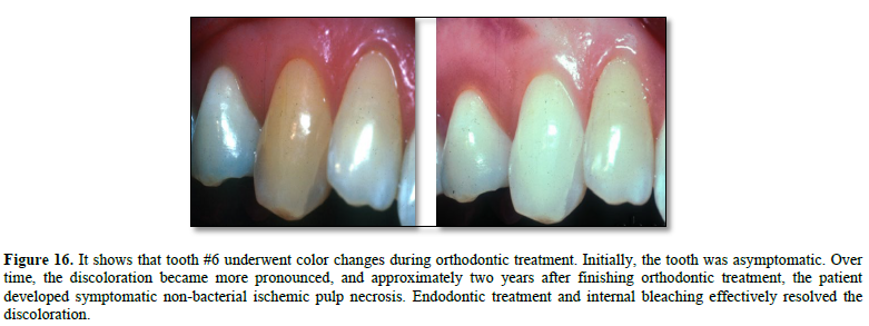
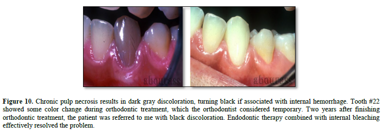
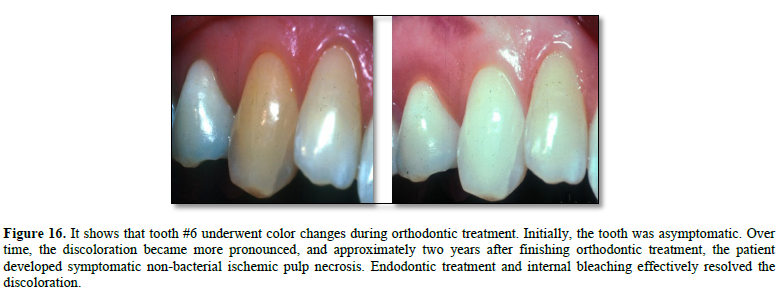
Beyond endodontics, tooth discoloration may happen during orthodontic treatment. While some consider it temporary, I have observed cases in which it leads to ischemic pulp necrosis and permanent staining. Discoloration can also occur in failing restorations, especially when functional, biological, and aesthetic criteria are not met.
4ROD Clinical Guideline:
Before performing endodontic treatment, examine all cleaned and exposed tooth surfaces for cracks, discoloration, abnormal wear, and anomalies.
Q7. You Introduced the ROTC Tests, the 3x3 Tooth-Crack Classification, and the EROIC Framework. How Do These Three Concepts Work Together to Enhance Crack Diagnosis?
Historically, the literature has predominantly emphasized root fractures and split-tooth conditions rather than common tooth cracks. Cameron addressed this in 1964, introducing the concept of cracked tooth syndrome into clinical dentistry. I categorize this as a line crack. Many line cracks remain undiagnosed, are mismanaged, or are frequently overlooked.
Additionally, the lack of standardized terminology, inconsistent classification, and dependence on unsupported, symptom-based flowcharts for managing tooth cracks have all led to widespread clinical uncertainty.
To highlight the importance of TSCs in the 4ROD diagnostic process, I will highlight key points from the master classes. This approach introduces an integrated model that aligns five core domains:
The above-listed content is available in the format of master classes available in the author’s Academy curriculum.
Q8. What is the 3x3 – Tooth Structure Cracks Classification Framework?
Tooth structure cracks are acquired pathological conditions with clinical implications, affecting pulpal, periapical, and periodontal health, as well as existing restorative and endodontic treatments.
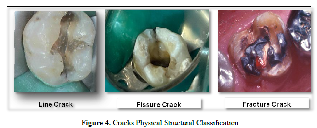
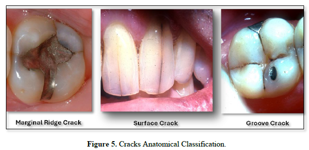
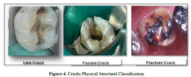
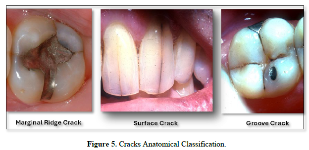
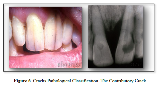
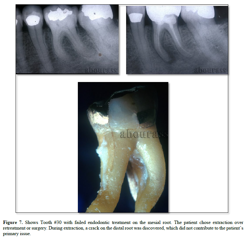
The 3 × 3 Tooth Structure Crack Framework is a practical, clinically oriented system for classifying tooth cracks based on their physical structure (Figure 4), anatomical location (Figure 5), and pathological effects (Figure 6).
This classification elevates tooth structure cracks from mere technical issues to a disease framework with significant pathological implications that are often overlooked in the literature.
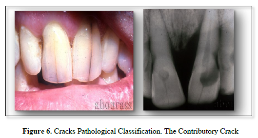
Teeth #8 and 9 each have a contributory line crack, causing severe bone loss, pulp necrosis, and grade 2 mobility in Tooth #9. The crack in tooth #8 caused pulpal calcification and an apical periodontitis.
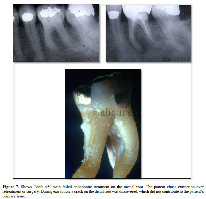
The Non-Contributory Crack
Q9. What is the EROIC Framework and How Does It Work?
EROIC is an acronym that stands for Endodontics, Restorations, and Occlusion-Induced Cracks.
The EROIC Etiology Framework is an evidence-based, clinically grounded model that explains the causes of tooth structure cracks. EROIC categorizes their origins into three interconnected etiologic domains: endodontic, restorative, and occlusion.
The framework analyzes clinical procedures in each domain using research and clinical evidence. Within the EROIC framework, the roots of these cracks are identified in endodontics, which is why it starts with 'E'. Evidence from 20 years of research by Professor Hagay Shemesh and his group at the University of Amsterdam, and others, shows that endodontic procedures can induce structural defects in the root canal walls, which serve as the initial point for microcrack development.
I have personally relied on Professor Hagay Shemesh’s research as my main reference for understanding tooth structure cracks from an endodontic perspective. By combining insights from restorative dentistry, occlusal considerations, and surgical and non-surgical observations, I developed the EROIC concept.
For instance, during exploratory surgery, a 3mm fissure was found on the buccal surface of the root's apical third. The EROIC analysis showed that the canal on the buccal side was overprepared. The pressure from the condensation of the root canal filling exceeded the strength of the canal walls, causing a burst and fissure in the thin apical third walls.
The clinician can use the EROIC framework to understand the mechanics behind crack formation. This involves examining factors such as the cavity preparation size, design, line angles, and the patient’s occlusal conditions.
Tooth structure cracks typically develop gradually and do not appear suddenly. They usually go through five stages over time, with progression influenced by factors like dental treatments, occlusal conditions, and masticatory function.
Q10. What Are The Five Stages Involved in the Development of a Tooth Crack?
When examining all elements involved in crack formation, it becomes clear that tooth cracks develop gradually rather than suddenly. Vertical and oblique root fractures, along with split-tooth conditions, are not caused by single impacts; instead, they represent the final stages of a prolonged process that begins earlier and progresses through five distinct stages.
The process begins with a mechanical action that generates a focused, localized stress either at the crown's axial-cavity floor line or along the root canal wall. Such initiating stress events include sharp line angles in the cavity, bypassing a root canal ledge or fractured instrument, preparing the post space, heavy occlusal loading, or parafunctional clenching.
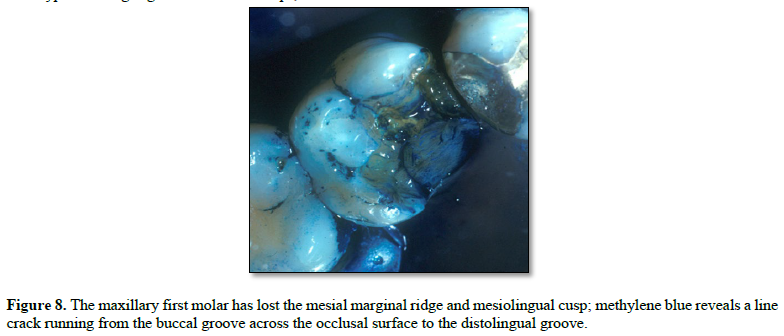
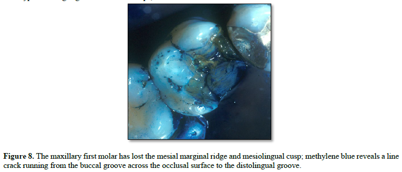
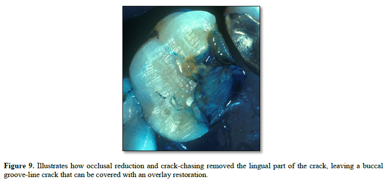
Microcracks form when local stress surpasses the tooth's strength, leading to tiny defects in the canal wall, line angle, or root surface. These defects are covered by restorations or root canal fillings. Clear visualization of the cracks requires coronal clean-out, fiber-optic transillumination (FOTI), and magnification.
A microcrack can serve as a stress concentration point. Over time, sustained stress causes the microcrack to grow and extend, eventually forming a visible line crack. Procedures such as endodontic retreatment, post placement, repeated functional loading, occlusal forces, structural loss, or additional restorative interventions can intensify mechanical stress on the microcrack, leading to the formation of the line crack, often observed in everyday practice.
With ongoing or repeated loading, the line crack can extend into the root or crown structure. Its path, depth, and growth rate are influenced by factors such as tooth anatomy, dentin structure, residual structural strength, restorations, occlusal forces, and the magnitude and direction of these forces.
A crack starting inside the tooth may grow towards the crown, root, vertically, or diagonally, depending on the loading pattern and remaining tooth structure.
The line crack eventually becomes visible in the clinical crown through visual inspection or coronal cleanout, and in the root during exploratory surgery. The specific nature, location, and impact of the crack on the tooth depend on factors like the remaining tooth structure thickness, the amount of applied stress, occlusal forces, and bacterial activity. Based on these factors, the defect or microcrack can grow and expand, appearing clinically as a line crack, fissure crack, or fracture crack.
4ROD Clinical Guideline:
Tooth Structure Cracks develop progressively through five stages: Stress Event, Initiation, Amplification, Propagation, and Manifestation.
Q11. What is the Purpose of the ROTC Framework?
The acronym ROTC stands for Rule-Out Tooth Crack. It is a diagnostic framework, a biologically based approach that employs optical, chemical, and mechanical techniques to detect and categorize cracks accurately.
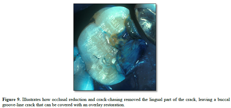
I developed the ROTC to help dentists routinely integrate crack detection into dental examinations. When clinicians actively look for cracks, they are more likely to identify them, given their widespread occurrence—particularly in both restored and intact teeth. Early detection enables management before cracks become symptomatic or progress to fissures and fractures.
ROTC has two main roles: detecting cracks and classifying their types. During regular dental check-ups, if no cracks are found, the dentist can continue with restorative or endodontic treatments. If a crack is discovered, ROTC offers tests to identify whether it is a line crack, fissure crack, or fracture crack. The tests include:
ROTC simplifies crack diagnosis, making it a clear and clinically measurable process. I see it as a valuable preventive diagnostic tool and hope it becomes a standard part of every dental appointment.
4ROD Clinical Guideline:
Before any dental treatment, rule out tooth structure cracks using ROTC tests
Q12. What is the Diagnostic Significance of Tooth Discoloration?
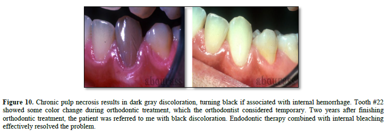
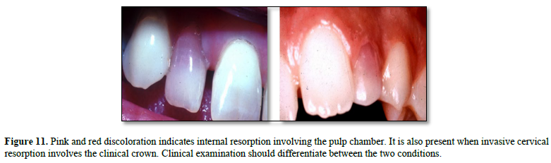
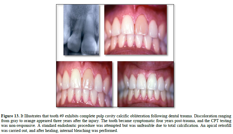
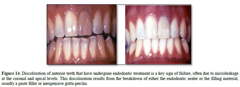
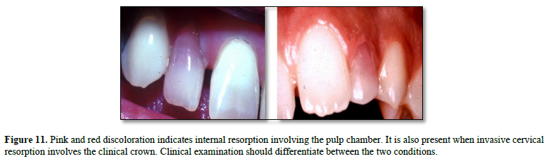
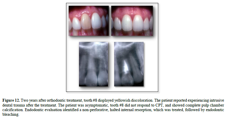
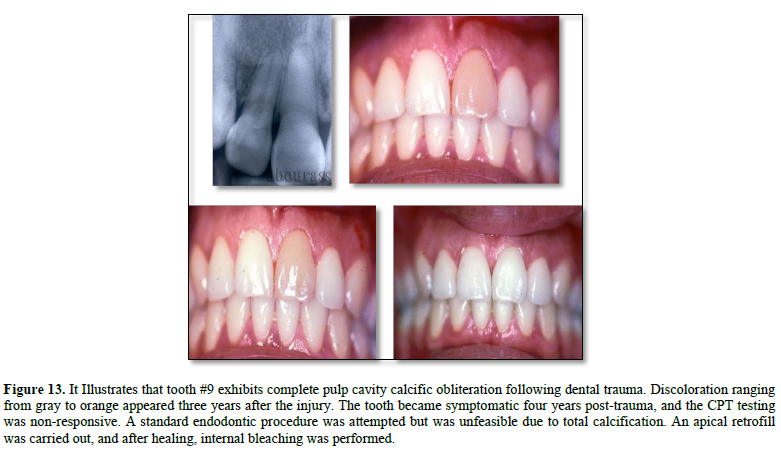
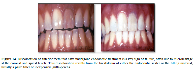
Tooth discoloration is more than a cosmetic concern; Color changes may indicate pulpal and periapical disease, failed root canal treatment, root resorption, or restoration issues. Discoloration can also affect self-esteem and social interactions, especially when multiple teeth are involved. Because it often develops gradually, it should be carefully evaluated as a diagnostic clue, even in the absence of symptoms.
Endodontic Causes: Pulp Necrosis
Endodontic Causes: Internal Resorption
Endodontic Causes: Pulp Necrosis with Root Resorption
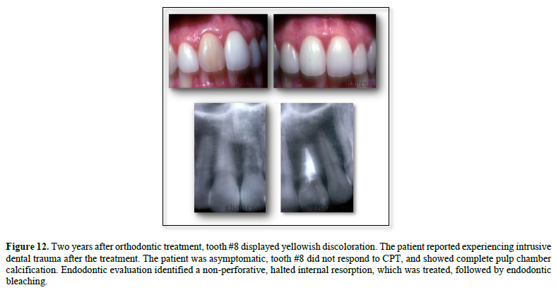
Endodontic Causes: Pulp Calcification
Endodontic Causes: Treatment Failure
Restorative Causes
Endodontics-Orthodontics Causes
4ROD Clinical Guideline:
Single-tooth discoloration is a preventable and treatable clinical issue rooted in endodontics. It can be effectively managed through standardized endodontic procedures, endodontic bleaching, and appropriate restorations.
Q13. How Can Dental Erosion Influence Pulpal and Endodontic Diagnosis?
Dental Erosion is a chemical-mechanical process that causes slow, irreversible loss of dental hard tissues. Unlike caries, which is bacteria-related, erosion results from repeated acid exposure and may progress unnoticed in its early stages. Its increasing prevalence is linked to modern dietary and lifestyle factors, reduced salivary flow, medications, GERD, chronic vomiting, eating disorders such as anorexia and bulimia, and alcoholism. In relation to this question, the focus will be on the pulpal and endodontic consequences of dental erosion.
Occlusion Implications
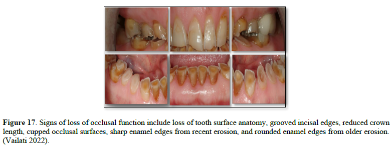
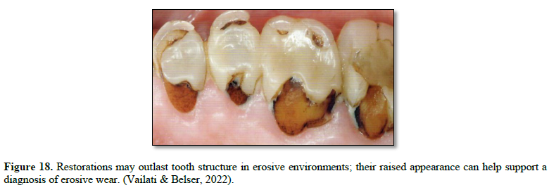
Endodontic Implications
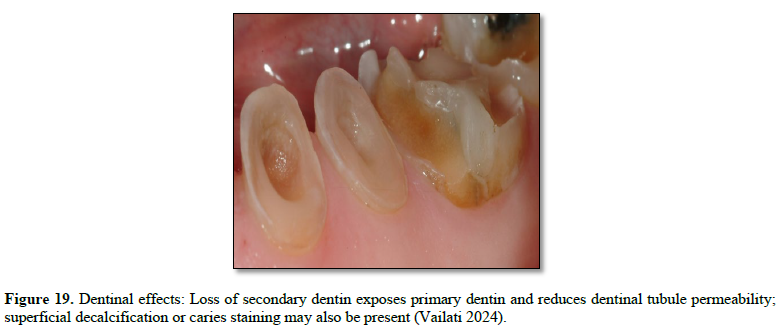
Pulpal Implications
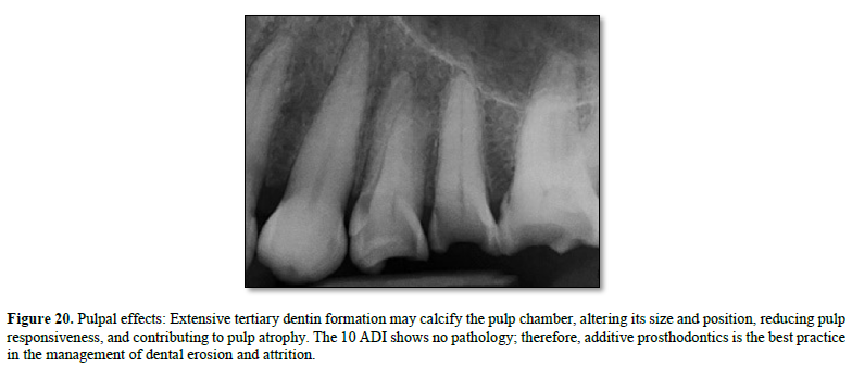
I would like to end my response with a personal note that I believe is very relevant to the clinical management of dental erosion and attrition.
My interest in tooth surface wear caused by erosion and attrition began during my advanced prosthodontics training. Back then, treatments mainly consisted of traditional crown-and-bridge prosthodontics and occlusal rehabilitation. While developing the curriculum on erosion and attrition, I encountered the work of Vailati and Belser from 2007, as well as their 2022 book, 3-Step-Additive Prosthodontics. I personally follow their principles and approach, considering them a pioneering method for managing severe dental erosion and attrition within prosthodontics in the academic program. Their work offers very detailed and valuable insights about Dr. Vailati’s contributions.
Q14. How Can Dental Attrition Influence Pulpal and Dentinal Health and Endodontic Diagnosis?
Physiologic dental attrition typically occurs with advancing age. However, in young patients, it is considered abnormal and often pathological; the primary cause is mechanical wear of the tooth structure. Causes include teeth grinding and clenching, malocclusion, and insufficient posterior support, all of which lead to uneven wear. Additionally, factors such as eating abrasive foods, chewing betel nut, tobacco use, and nail biting can worsen attrition, especially when natural teeth oppose ceramic restorations.
Dentition Effects
Advanced dental attrition is a significant issue that causes several problems, including a decrease in the Occlusal Vertical Dimension (OVD), possible super-eruption of posterior teeth, reduced resistance to fracture, occlusal instability, loss of masticatory function, and a unilateral cusp-to-cusp relationship.
Occlusal Effects
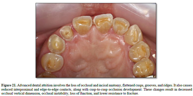
Pulp's Response to Dental Attrition
The literature indicates that histologically, dental attrition leads to the formation of irregular secondary dentin directly beneath the worn dentin. The odontoblast layer flattens, and there is increased cellularity below the irregular secondary dentin, indicating local pulpal responses. The pulp exhibits increased vascularity and cellular activity, along with mild inflammation and circulatory issues. However, the overall structure of the pulp remains largely intact.
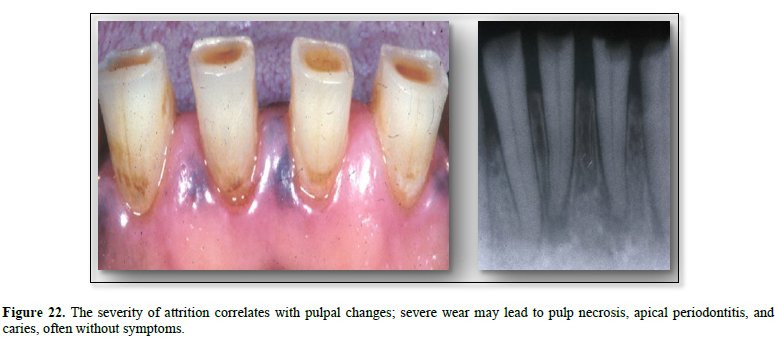
Q15. How Does Dental Abrasion Affect the Pulp?
Scientific studies highlight several risk factors for dental abrasion, such as an abnormal CEJ, cervical enamel defects, gingival recession, gingival dehiscence, an acidic diet, gastric reflux, bulimia, and chemicals used in teeth whitening.
In endodontics, dental abrasion is the main cause of dentinal hypersensitivity (DHS). Prevalence 7.5% in dental attrition and 71.9% with dental abrasion. Cervical abrasion exposes the thin cervical dentinal tubules on the buccal side of the CEJ. This direct exposure allows stimuli—such as thermal, chemical, or physical changes—to disturb the flow of dentinal fluid, leading to hypersensitivity symptoms. These symptoms include sharp, fleeting pain triggered by thermal changes, airflow, touch, osmotic substances, and chemicals. The pain occurs only in response to specific stimuli, not spontaneously or continuously, and can interfere with activities like eating, drinking, speaking, and brushing. It rarely affects the pulp or periapical tissues.
Q16. How Does Dental Abfraction Impact Pulpal and Dentinal Health?
Traditionally, the term abfraction is used to describe the buccal surface (CEJ) primarily, wedge-shaped 1-3mm depth cavities or defects with sharp line angles. Usually extends below the gingival margin at the cervical zone of the anterior and premolar teeth. The problem is observed on multiple misaligned teeth. The abfraction lesions are typically asymptomatic, and in advanced cases, they can be associated with mild sensitivity.
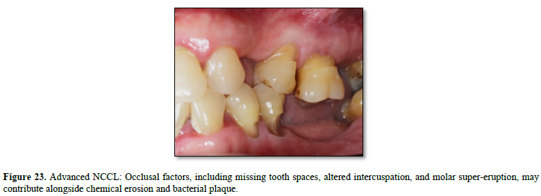
In terms of etiology, the literature focused on bruxism and parafunctional habits.
Over the past 2 decades, the term abfraction has been rephrased as Non-Carious Cervical Lesion (NCCL). Reportedly, this is due to the highly distinctive and harmful consequences of the observed defect, which affects multiple teeth at once. NCCLs are asymptomatic until the pulp is exposed, and in many cases, pulp exposure and necrosis occur without symptoms.
The literature emphasizes parafunctional activities such as clenching, grinding, nail biting, tongue thrusting, and chewing of oral tissues, all of which involve excessive vertical or horizontal force on the teeth. Other etiology theories are discussed in the presentation on the subject in the academic curriculum.
Q17. Which Developmental Anomalies That Have Significant Endodontic Implications?
The R4 discussion focuses on three anomalies: palatogingival groove (PGG), dens invaginatus (DI), and dens evaginatus (DE). Each will be presented separately because early recognition is essential to prevent pulp infection, localized periodontal breakdown, and complex, multidisciplinary complications.
Palatogingival Groove (PGG)
PGG occurs in about 2.9% to 4% of cases but can affect up to 30% of the Asian population, usually involving the maxillary lateral incisor. It often remains asymptomatic for years, but flare-ups can lead to serious endodontic and periodontal problems.
Gu 2007 classified PGG into three types based on groove extension. Type I features a groove limited to the coronal third with normal root canal anatomy. Type II shows a groove extending beyond the coronal third to the apex, maintaining a normal root canal. Type III involves a groove that extends beyond the coronal third, exhibiting complex C-shaped anatomy and additional canals; most are 6–10 mm long, though some reach the apex.
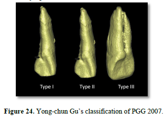 Treatment typically includes combined endodontic and periodontal approaches, such as curettage of affected tissues, saucerization to remove the groove, sealing the groove, and, if necessary, guided tissue regeneration. Some practitioners also consider intentional replantation as an option. In terms of management, referring to an endodontist is highly recommended.
Dens Invaginatus (DI)
Dens Invaginatus, also known as Dens Dente, is a rare developmental anomaly with a prevalence of 1-2%. A deep pit, a bulge, or a peg-shaped tooth characterizes it. The Invagination is open to the mouth; it traps saliva, food, and bacteria, which eventually lead to pulpal pathology. (Figure 25).
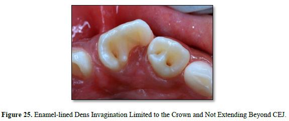
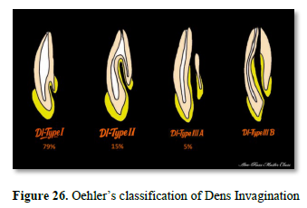
Oehler classification (Figure 26) indicates that DI cases exhibit many anatomical complexities, such as multiple canals, constrictions, isthmuses, and communication with the PDL, which make endodontic treatment very challenging. Furthermore, many DI cases may require surgical endodontics in addition to standard endodontic treatment. Therefore, referral to an endodontist is highly recommended.
Dens Evaginatus (DE)
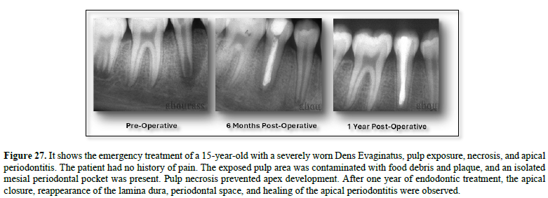
Dens Evaginatus is a developmental dental anomaly characterized by an extra cusp-like projection on the tooth's surface, often containing pulp extension or a pulp horn. It occurs in less than 1% of cases, primarily among Asian and Native American populations, especially in the lower premolars, and tends to affect both sides. When the affected tooth erupts and contacts opposing teeth, the protrusion is often fractured or worn down, exposing the pulp. This exposure stops tooth development, resulting in an open apex and an apical lesion (Figure 27). The management of these cases is comprehensively discussed in the academy curriculum, including literature-based case reports.
No Files Found


Internationally Accepted
Share Your Publication :