Menu
  • Publish Your Research/Review Articles in our High Quality Journal for just USD $99*+Taxes( *T&C Apply)

    Offer Ends On

Review Article

The Periodontal Ligament, Link Between the Root and Alveolar Bone

Michel Goldberg*, Victor Arana-Chavez E and Chen Zhi

Corresponding Author: Michel Goldberg, Department of Oral Biology, Faculty of Fundamental and Biomedical Sciences, INSERM UMR-S1124, Paris Cité University, France.

Received: March 14, 2023 ;    Revised: March 18, 2023 ;    Accepted: March 21, 2023 ;   Available Online: March 31, 2023

Citation: Goldberg M, Arana-Chavez VE & Zhi C. (2023) The Periodontal Ligament, Link Between the Root and Alveolar Bone. J Oral Health Dent Res, 3(1): 1-9.

Copyrights: ©2023 Goldberg M, Arana-Chavez VE & Zhi C. This is an open-access article distributed under the terms of the Creative Commons Attribution License, which permits unrestricted use, distribution, and reproduction in any medium, provided the original author and source are credited.

Share Your Publication :

Views & Citations

938

Likes & Shares

0


Global Views

  • Abstract
  • Full Text
  • Images
  • Tables
  • References
  • PDF
  • Supplementary Files

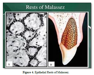
The periodontal ligament (PDL) connects the root and alveolar bone. Anchored to mineralized surfaces, it includes collagens and oxytalan fibers. Metalloproteinases play role in the turnover of type I collagen and extracellular matrix proteins. After dissociation of the epithelial sheath, epithelial rests of Malassez constitute a source of stem cells. Osteonectin and biglycan were detected in PDL cells. The levels of the receptor activator of NFkB ligand (RANKL) and osteoprotegerin (OPG) implicate changes of amount of RANKL, OPG, matrix proteins (collagen, fibronectin, SPARC, glycosaminoglycans (GAGs) and proteoglycans (PGs), MMPs and TIMP - 1 to 4). We explore here 1) the attachment function of the PDL (eruption, formation and supportive functions), 2) the implication of stem cells in healing, regulation of bone volume, and PDL regeneration, 3) the contribution of fibroblast-like and pericytes to periodontal inflammation, 4) sensory events (Ruffini-like mechanoreceptors), 5) vascular supply, and 6) because the cells of the PDL differentiate into cementoblasts and osteoblasts, this phenotypic availability contributes to heal the tooth supporting tissues. This structure initiates a process of self-organization resulting in a functional architecture of the PDL.

Keywords: Periodontal ligament, Radicular tooth, Alveolar bone, Cementum, Collagen fibers, Oxytalan fibers, Metalloproteinases, Epithelial rest of malassez


INTRODUCTION

The periodontal ligament acts as an attachment between the bone and dental root(s), including mostly collagens and oxytalan fibers.  Many terms have been used to describe the periodontal ligament, including desmodont, gomphosis, peri-cementum, dental periosteum, alveolo-dental ligament and periodontal membrane. The PDL is anisotropic, heterogeneous, viscoelastic and possesses non-linear elastic properties. The effects of the PDL are restricted to the maxillary and mandibular alveolar bone [1-4].

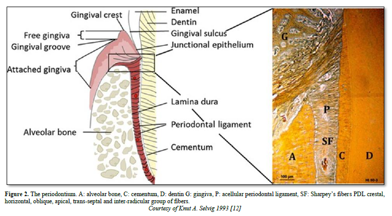
The periodontal ligament is thinner in the middle of the root and wider near the root apex. The “normal” width of the ligament (mean 25mm) is decreasing with age [5-8]. The functions of the ligament implicate nutrition, and multiaxial loading. The periodontal ligament consists of fibers coming from the pulp and gingiva, from the cribliform plate, from the vascular network, and from the nerves supply. Alveolar bone and cement are mineralized whereas the periodontal tissue includes the ligament and the gingival lamina propria. The PDL is composed by clastic cells, loose connective tissue (collagens and oxytalan fibers), fibroblasts and cell rests of Malassez [9] (Figure 1).

Characterization of periodontal ligament cells in vitro [10-11]

Alveolar bone can be divided into two main parts

  • A thin layer of compact bone (the cortex of the alveolus) lining the alveolus proper in which Sharpey’s fibers insert (lamina dura)
  • The main alveolar bone surrounding the lamina dura. The alveolar bone remains spongy and porous.

In addition to the two layers, dense compact and cancellous bones form another layer, lining the alveolar socket. Blood vessels and nerves perforate the inner cribliform plate.

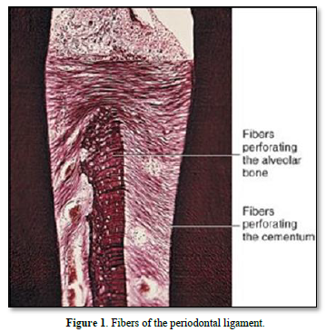
Periodontal ligament comprises five different groups of fibers mostly formed by fibroblasts:

  • In multiple rooted teeth: alveolar crest, horizontal, oblique, apical, and inter-radicular fibers
  • In single rooted teeth, groups of fibers are found forming the dento-gingival, alveolo-gingival, circular, and dento-periosteal group of fibers [12].

The principal fibers ended from part and other in the cementum and in bone (named in such case Sharpey fibers) (Figure 2).

Composition of the periodontal ligament

Functions

Formative function plays a critical role in periodontal repair and regeneration, with high turnover rate of the PDL collagen.

Nutritive function.

Proprioceptive function.

Supportive function inside the bonny socket.

Homeostatic function.

  • Tensional theory: PDL transmits the force to bone and elastic deformation of the socket.
  • Viscoelastic theory: the extracellular forces are pulsed from the periodontal ligament into marrow spaces through the cribliform plate.

The PDL is formed by:

  • Fibroblasts implicated in the synthesis and secretion of collagens.

Collagens

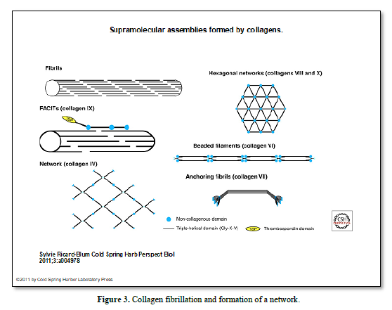
The collagen containing connective tissue includes type I, III, and VI and Type XII collagens (53-57%), embedded in the intercellular substance. Collagen fibers have roughly 55 nm in diameter (0,15 to 0,38 mm) [6-10]. Water has been estimated to be 70% (Figure 3).

  • Type I collagen is moderate in the periodontal ligament, and weaker in bone and cementum. Linked covalently to type I collagen, these fibers contain a high content of cysteine. Type I collagen is found in the periphery of Sharpey’s fibers, forming an attachment to alveolar bone, and around nerves and blood vessels.
  • Type III collagen is formed by 3 identical alpha 1 chains with a high content in hydroxyproline, and low in hydroxylysine.
  • Type V collagen is present in small amount in the periodontal ligament.
  • Type VI collagen is part of the oxytalan fibers system. Staining for type VI collagen was strong whereas predentin and dentin were negative.
  • Type XII collagen is present at pressure sites.

Collagen type XIV is expressed in the PDL, forming bundles 5 micrometers in diameter.

  • Three major groups of integrins have been reported in the PDL:
  1. I-domain-containing collagen-binding integrins
  2. Non-I-Domain integrins linked via ‘bridging’ molecules to collagen
  • Transforming growth factor - activating integrins [11-13].

In vertebrates, the integrin family is composed of 18 subunits and 8 subunits that may assemble into 24 different heterodimers.

  • Three types of elastic fibers are found in the PDL: elastin, oxytalan and elaunin.
  • Oxytalan is an immature form of elastin. It constitutes 3% of the extracellular fibers’ composition. Naturally elastic, the fibers run both parallel and oblique to the root surface. They contain reticulin and elastin. These fibers, 5-2.5 micrometers in diameter, are oval in shape. They are tooth support, and contribute to fibroblast migration.
  • Elaunin contains only a small amount of elastin.

PA-TCH-SP and Con A staining of carbohydrates are very useful in identifying oxytalan fibers at the ultrastructural level [15]. They consist of bundles of filaments approximately 150 Å in diameter with an interfilamentous substance of the same diameter.

  • Ground substance
  • Glycosaminoglycans and proteoglycans: Glycosaminoglycans (GAGs) and proteoglycans (PGs)
  • Hyaluronan: play role in tissue permeability, and cell mobility. CS /DS hybrids
  • Decorin & biglycan, and heparin sulfate are components of the ground substance. CSPGs are among major PGs of the PDL. The components include also fibronectin, tenascin, and vitronectin
  • Fibromodulin and lumican are KSPGs, modulating collagen fibers formation.
  • Periostin, initially found in the periosteum, favor osteoblast attachment and spreading, and interact with collagen Type I.
  • CD44 known as lymphocyte homing receptor is a trans-membrane glycoprotein, associated with actin microfilaments, binding to fibronectin, laminin, and collagen.
  • Glycoproteins: Nidogen, vitronectin, tenacin, and thrombospondin have been identified in the periodontal ligament.
    • CD44 known as lymphocyte homing receptor is a trans-membrane glycoprotein, associated with actin microfilaments, identified in the periodontal fibroblasts.
  • Fibroblastic cells are involved in collagen degradation. At early passage, PDL cells exhibits contact inhibition. Periodontal ligament cells exhibit significantly distinct characteristics.
  • Cementoblasts are forming acellular and cellular cementum.
  • Osteoblasts: are cuboidal cells loaded by microfilaments. Tight junctions form a transport system in bone (syncytium). Osteoblasts, under the influence of osteotropic hormones, produce MMPs which appear to remove soft tissue that precludes access of osteoclasts to the mineralized tissue surface [16-19].
  • Osteoclasts/cementoclasts Although there is strong evidence for the involvement of MMPs in the resorption of bone and in the inflammation-mediated destruction of periodontal tissues, the role of MMPs in the remodeling of mature soft connective tissues remains equivocal.
  • Epithelial rests of Malassez have slow turnover [20,21]. The epithelial cell rests of Malassez, rather than being ‘cell rests’ are an important source of stem cells that might play a pivotal role in periodontal regeneration. They also have the capacity to differentiate into a mesenchymal phenotype and thus represent a unique stem cell population within the periodontal ligament (Figure 4).

Cells of the PDL

  • Defense cells such as monocytes/macrophages constitute 4% of the cell population in PDL. Mast cells eosinophils are involved in phagocytosis. Neutrophils and lymphocytes develop in case of inflammation. They are close to cementum.
  • Blood vessels: the volume of the lumens vary between 10-30% in the PDL. Fenestrations play role in capillaries.
  • Nerve: sensory and myelinated sensory have mensuration varying between 5 mm and 15mm. Unmyelinated nerves are both sensory and autonomic (0.5 mm).
  • Mechanoreceptors Ruffini-like mechanoreceptor A are reactive for cytochrome oxidase activity and acts as neuropeptide Y.
  • Terminal Schwann cells have both non-specific cholinesterase and acid phosphatase activities.
  • The function of epithelial rests of Malassez seems to be their involvement in repair and regeneration. They possibly act as ankylosis inhibitors. They have been implicated in the development of periodontal pockets. They express ameloblastin that play a role in the differentiation of ameloblasts and induction of cementoblasts.

Mensuration of the PDL The “normal” width of the ligament (mean 25mm) is decreasing with age. The thinnest part of the PDL is located in the middle third of the root. It increases near the apex.

Periodontal Ligament composition [22-23]:

  • Water 70%
  • Proteins including the cells associated with innervation, vascularization, and extracellular matrix molecules. The ligament presented a high density of collagen fibrils grouped in bundles and oxytalan fibrils. Fibroblast-like cells are characterized by collagen production and possess some other osteoblastic features. They also produce cytokines and chemokines in response to inflammation promoters.
  • Cells of the periodontal ligament include fibroblasts (playing role in homeostasis and regeneration). They are ecto-mesenchymal in origin. They are spindle-shaped. Fibroblasts are fusiform with many cytoplasmic processes. They are mobile and contractile. TGF-beta promotes tissue matrix formation by stimulating both the synthesis of matrix proteins They contain large amount of RER and a well-developed Golgi apparatus (collagen, fibronectin and SPARC), metalloproteinases (MMPs), proteinase inhibitors (TIMP-1 to -4) and by decreasing the synthesis of MMPs, except the 72 kDa-gelatinase (gelatinase A). They exhibit osteoblastic properties (alkaline phosphatase activity, responsiveness to parathyroid hormone, production of bone sialoprotieins in response to 1,25 dihydroxyvitamin D).
  • Fibroblasts possess receptor activator of NF-kappa B ligand (RANKL) and osteoprotegerin (OPG), both playing a key role in bone metabolism. On-stimulation with bacterial lipopolysaccharides, PDL fibroblasts up-regulate transcription of various cytokines and chemokines. Effects of growth factors/cytokines include proteins or steroid hormones (BMP-2 and -7, PTH, IGF, vit D) known as signaling molecules and stimulating cell proliferation and differentiation. They bind to cellular receptors. Fibroblasts are fusiform with many cytoplasmic processes. They are mobile and contractile. TGF-beta promotes tissue matrix formation by stimulating both the synthesis of matrix proteins (collagen, fibronectin and SPARC), metalloproteinases (MMPs), proteinase inhibitors (TIMP-1 to -4) and by decreasing the synthesis of MMPs.
  • FGF2 is a heparin-binding cytokine.
  • TGF is member of a superfamily of growth factors with multifunctional effects. Three isoforms i.e., TGF 1, 2 and 3 have been detected.
  • PDGF platelets produce and release growth factors involved in angiogenesis, inflammation and immune response which enhance tissue repair. They are actively involved in tissue regeneration and wound healing.
  • Osteoblasts are found at the surface of alveolar bone. They are connected one-to-another via gap junctions and desmosomes. They contact osteocytes.
  • Osteoclasts or bone resorption cells, play role in bone remodeling. Multinucleated cells with a ruffle border adjacent to the resorbing surface enclosed by a clear zone. Numerous mitochondria suggest high metabolic activity. Initially, the bone is demineralized and then, the exposed matrix is degraded. Acting on cementum or dentin surfaces, cementoclasts or dentinoclasts are referred respectively.
  • Defense cells, and undifferentiated mesenchymal cells gave a PGE2- and isoproterenol-mediated cAMP response, but did not respond in similar fashion to calcitonin or PTH. The periodontal ligament cells produce a bone-associated protein, osteonectin. In addition, osteonectin and biglycan were detected in the cells. Cells of the periodontal ligament include fibroblasts (homeostasis and regeneration). They are ecto-mesenchymal in origin. They are spindle-shaped. They contain large amount of RER and a well-developed Golgi apparatus. They exhibit osteoblastic properties (alkaline phosphatase activity, responsiveness to parathyroid hormone, production of bone sialoproteins in response to 1,25 dihydroxyvitamin D. Fibroblasts possess receptor activator of NF-kappa B ligand (RANKL) and osteoprotegerin (OPG), both playing a key role in bone metabolism. On-stimulation with bacterial lipopolysaccharides, PDL fibroblasts up-regulate transcription of various cytokines and chemokines.
  • Proteins synthetized by the periodontal ligament [22-23].
  • Extracellular matrix proteins (collagens-namely type I, type III and XII), oxytalan fibers, bone sialoprotein, DMP-1, dentin sialoprotein, fibronectin, osteonectin, osteopontin, tenascin), and the ‘Secreted Proteins Acidic and Rich in Cysteine’ (SPARC), elastic fibers (elastin, oxytalan, and elaunin), metalloproteinases (MMPs) and tissue inhibitors of metalloproteinases (TIMP- 1 to - 4) that are structural components of the PDL [19]. Alkaline phosphatase, growth factors (IGF, TGF and platelet-derived growth factor).
  • Transcription factors Runx-2, also known as Cbfa1, and osterisx.
  • PDLSCs express common mesenchymal stem cell (MSC) surface markers and some pericyte markers such as STRO-1, STRO-4, CD29, CD73, CD90 (Thy1), CD106 (VCAM-1), and CD146 (MUC18). They are negative for the expression of endothelial (CD31), hematopoietic (CD14, CD34, CD45, CD79a), and helper immune antigens (CD11b, CD19, HLA-DR, CD40, CD54, CD80, CD86). PDLSCs located in the perivascular wall of periodontal ligament. PDLSCs are a subpopulation of MSCs located in the perivascular space which share similarities with pericytes.
  • Following culture of the cells for 3 days, high glucose media induced the expression of NANOG, octamer-binding transcription factor 4, (sex determining region Y)-box 2, cluster of differentiation 166 (CD166), periostin and β-catenin. After 14 days in high glucose condition, alkaline phosphatase activity increases. A high number of calcified nodules were formed on day 28. These results suggest that high glucose induced bone formation by elevating the expression of stem cell markers, particularly CD166, and this induction may be regulated through β‑catenin [26].
  • Matrix metalloproteinases (MMPs), also known as matrixins, are calcium-dependent, zinc-containing endopeptidases. Other family members are adamalysins, serralysins, and astacins. The MMPs belong to a larger family of proteases known as the metzincin superfamily.
  • Synthetized in a latent form (zymogen), they are secreted as proenzymes and require extracellular activation. They are inhibited by tissue inhibitors of metalloproteinases (TIMP-1 to TIMP-4) [16,17]. They are grouped into collagenases, gelatinases, stromelysins, et matrilysins. 8 classes are structural (5 classes are secreted) and 3 classes are of the membranes type (MT-MMPs).
  • The MMPs include
    • One peptide signal or pre-domain
    • One pro-domain, with proteolytique cleavage, mandatory for the enzyme activation
    • A catalytic domain with zinc fixation
    • One hemopexin-like domain (Figure 5)

Innervation

Branches of the Vth cranio-facial nerve arises from the trigeminal nerve through its superior or inferior alveolar branches. The average density of myelinated nerve fibers increased by arriving closer to the apex. The diameter of myelinated fibers varied between 5.3 and 7.8 μm. The innervation is close to the alveolar bone. Isolated myelinated axons showed a tendency to group around large blood vessels. Dense innervations by myelinated nerve fibers reveals that apical as well as mesial and buccal sites are more densely innervated [11].

Type I receptor received its blood supply by diffusion whereas Type II receptor had a direct blood supply.  Under each region Ruffini terminals and smaller terminals resembling free nerve endings were observed. The Ruffini terminals were unencapsulated and the majority had diameters of 2-3 microns. The terminals were observed near the junction of the inner and middle zones of the periodontal ligament with the axons running from the alveolar aspect. Periodontal mechanoreceptors, even those with more rapidly adapting properties, are Ruffini terminals.

Type II mechanoreceptors are the primary mechanoreceptors in the periodontal ligament. The periodontal Ruffini endings display dendritic ramifications with expanded terminal buttons and furthermore, are ultrastructurally characterized by expanded axon terminals filled with many mitochondria and by an association with terminal or lamellar Schwann cells.

Corpuscles of Ruffini are present in the PDL, forming a peripheral innervation. The blood capillaries showed a continuous endothelium. They are showing little adaptation capacities. They respond to sustained pressure and they are slowly adapting receptors.

Histochemically, the axon terminals are reactive for cytochrome oxidase activity, and the terminal Schwann cells have both non-specific cholinesterase and acid phosphatase activity. It has been suggested that the Ruffini endings have a high potential for neuroplasticity. Immunoreactivity for p75-NGFR (low-affinity nerve growth factor receptor) and GAP-43 (growth-associated protein-43), both of which play important roles in nerve regeneration/development processes, have been reported in the periodontal Ruffini endings. In experimental studies on nerve injury to the inferior alveolar nerve, the degeneration of Ruffini endings takes place immediately after nerve injury, with regeneration beginning from 3 to 5 days later, and the distribution and terminal morphology returning to almost normal at around 14 days. During regeneration, some regenerating Ruffini endings expressed neuropeptide Y. On the other hand, the periodontal Ruffini endings show stage-specific configurations which are closely related to tooth eruption, suggesting that mechanical stimuli due to tooth eruption and occlusion are a prerequisite for the differentiation and maturation of the periodontal Ruffini endings.

Numerous myelinated and non-myelinated nerve fibrils, as well as nerve endings and mechano-receptors are identified in the PDL [23]. Using a PGP 9.5 antibody staining it was shown that the apical region was richly supplied with nerve terminals. Neurofilament protein (NFP) and glia-specific S-100 protein., released by sympathetic nerves have been identified in the ligament, whereas there is no evidence of a parasympathetic innervation in the PDL [22-24].
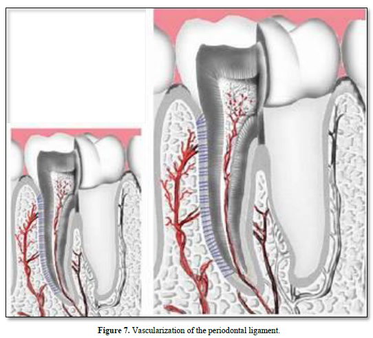
Vascular supply

Vascularization arises from the superior and inferior alveolar arteries, derived from a series of perforation arteries that pass through the alveolar bone, and occupy interstitial spaces. Many arteriovenous anastomoses occur within the PDL. Lymphatic vessels tend to follow the venous drainage. The abundance of microfilaments and microtubules as well as the presence of intermediary and tight junctions characterize the vascular supply.

Vascular specific markers CD31 and vascular endothelial growth factor (VEGFA) are highly expressed in the PDL. SMAD 3, integrins and VEGF have increased expression in the PDL during orthodontic tooth movement. Nexus between the cell processes indicate that these cells are contractile and motile. The vascular distribution is asymmetric: vascularization is seen to be adjacent to bone, but not to cementum [25].

Arteries enter the PDL space from the alveolar bone. They are referred to as perforating arteries.  Dental inter-septal artery enters the PDL space and contact anastomosis with gingiva blood vessels. They are referred to as “hydraulic pressure distribution” or “functional regeneration and adaptation”.  Neuropeptides are released and fenestrations allows nutriments to diffuse in the PDL. The veins drain into the interdental veins or into the periapical plexus.

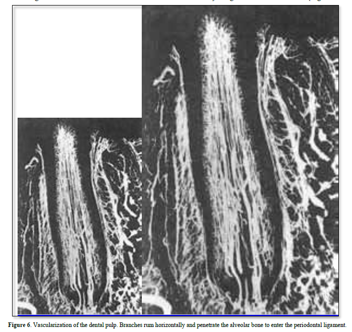
The lymphatic vessels drain into the regional lymph nodes, and finally merge with the thoracic duct (Figures 6 & 7).

CONCLUSION

The periodontal ligament constitutes a physical link between the cementum and bone of the alveolar crest. In addition to the PDL functions as support, sensory, nutritive, and remodeling, an attachment is induced to the inner bone socket. Epithelial Hertwig epithelial rests may provide stem cells, implicated in the healing and regeneration of the PDL. Tissue repair of the ligament, and sensory events are requiring nerve corpuscles (namely Ruffini endings) and vascularization. PDL include collagen and oxytalan fibers, phosphorylated and non-phosphorylated molecules, blood serum derived alpha2-HS-glycoprotein, fibronectin, glycosaminoglycans and proteoglycans, a number of extracellular matrix proteins (including fibronectin, and SPARC proteins), metalloproteinases and tissue inhibitors of MMPs [26]. This heterogeneous composition shed lights on the complexity of this structure and to the diversity of functions.

It is concluded that PDL has 1) an attachment function to bone and cementum (eruption, formation and supportive functions), 2) STEM cells are implicated in healing. The PDL is involved in the regulation of bone volume, PDL repair, tissue homeostasis and regeneration, 3) fibroblast-like and pericytes cells of the PDL contribute to periodontal inflammation. Hence, the cells of the PDL contribute and release molecules implicated in periodontal inflammation, 4) they are also implicated in sensory events requiring nerve corpuscles, and providing vascular supply and nutrients to the cementum, alveolar bone and to the PDL itself. 5) because the cells of the PDL may differentiate into cementoblasts and osteoblasts their phenotypic availability contributes to the healing and regeneration of tooth supporting tissues [27].

  1. Beertsen W, Mcculloch CA, Sodek J (1997) The periodontal ligament: A unique, multifunctional connective tissue. Periodontology 13: 20-40.
  2. Nanci A, Bosshardt DD (2000) Structure of periodontal tissues in health and disease. Periodontology 40: 11-28.
  3. Bosshardt DD, Selvig KA (1997) Dental cementum: The dynamic tissue covering of the root. Periodontology 13: 41-75.
  4. Panagiotopoulou O, Kupczik K, Cobb SN (2011) The mechanical function of the periodontal ligament in the macaque mandible: A validation and sensitivity study using finite element analysis. J Anatomy 218(1): 75-86.
  5. Mortazavi H, Baharvand M (2016) Review of common conditions associated with periodontal ligament widening. Imag Sci Denti 46(4): 229-237.
  6. Berkovitz KB, Moxham BJ, Newman HN (1995) The periodontal ligament in health and disease Mosby-Woljfe.
  7. Berkovitz BK (2004) Periodontal Ligament: Structural and clinical correlates. Dent Uptake 31(1): 46-50.
  8. Berkovitz BK, Holland GR, Moxham B (2018) The periodontal ligament in health and disease. Oral Anatomy, histology and embryology. 5th Elsevier London.
  9. Zeichner-David M (2006) Regeneration of periodontal tissue. Periodontology 41: 196-217.
  10. Ricard-Blum S (2011) The collagen family. Cold Spring Harb Perspect Biol 3: a004978.
  11. Somerman MJ, Young MF, Foster RA, Moehring JM, Imm G, et al. (1995) Characteristics of human periodontal ligament cells in vitro. Archs Oral Biol 35(3): 241-247.
  12. Selvig KA (1991) Structure and function of the periodontium. Dent Update 18(7):292-297.
  13. Barczyk M, Bolstad AI, Gullberg D (2013) Role of integrins in the periodontal ligament: Organizers and facilitators. Periodontology 63(1): 29-47.
  14. Takagi M, Parmley RT, Yagasaki H, Toda Y (1984) Ultrastructural cytochemistry of oxytalan fibers in the periodontal ligament and microfibrils in the aorta with the periodic acid-thiocarbohydrazide-silver proteinate method. J Oral Pathol 13(6): 671-678.
  15. Takagi M, Yagasaki H, Baba T, Baba H (1985) Ultrastructural visualization of carbohydrates in oxytalan fibers in monkey periodontal ligaments J Histochem Cytochem 33(10): 1007-1014.
  16. Arpino V, Brock M, Gill SE (2015) The role of TIMPs in regulation of extracellular matrix proteolysis. Matrix Biol 44-46 :247-254.
  17. Behm C, Nemec M, Weissinger F, Rausch MA, Andrukhov O, et al. (2021) MMPs and TIMPs expression levels in the periodontal ligament during orthodontic tooth movement: A systematic review of in vitro and in vivo Int J Mol Sci 22(13): 6967.
  18. Sodek J, Overall CM (1992) Matrix metalloproteinases in periodontal remodeling. Matrix Suppl 1: 352-362.
  19. Decup F, Six N, Palmier B, Buch D, Lasfargues J-J, et al. (2000) Bone sialoprotein-induced reparative dentinogenesis in the pulp of rat’s molar. Clin Oral Investig 4(2): 110-119.
  20. Rincon JC, Young WG, Bartold PM (2006) The epithelial rests of Malassez- a role in periodontal regeneration? J Periodontal Res 41(4): 245-252.
  21. Xiong J, Gronthos S, Bartold PM (2013) Role of epithelial cell rests of Malassez in the development, maintenance and regeneration of periodontal ligament tissues. Periodontology 63(1): 217-233.
  22. Huang Y, Corpas LS, Martens W, Jacobs R, Lambrichts I (2011) Histomorphological study of myelinated nerve fibres in the periodontal ligament of human canine. Acta Odontol Scand 69(5): 279-286.
  23. Dean R (2017) The periodontal ligament: development, Anatomy and function. OHIDM 16: 1-7.
  24. Maeda T, Ochi K, Nakakura-Ohshima K, Youn SH, Wakisaka S (1999) The Ruffini ending as the primary mechanoreceptor in the periodontal ligament: Its morphology, cytochemical features, regeneration, and development. Crit Rev Oral Biol Med 10(3): 307-327.
  25. Frank R, Fellinger E, Steuer P (1976) Ultrastructure of the alveolodental ligament of the rat. J Biol Bucale 4(4): 295-313.
  26. Rooker SM, Liu B, Helms JA (2010) The role of Wnt signaling in the biology of the peridontium. Dev. Dyn 239(1): 130-147.
  27. Seubbuk S, Sritanaudomchai H, Kasetsuwan J, Surarit R (2017) High glucose promote the osteogenic differentiation capability of human periodontal ligament fibroblasts. Mol Med Rep 15: 2788-2794.

    No Files Found