
-
Publish Your Research/Review Articles in our High Quality Journal for just USD $99*+Taxes( *T&C Apply)
Offer Ends On
Publish Your Research/Review Articles in our High Quality Journal for just USD $99*+Taxes( *T&C Apply)
Offer Ends On
Manisha Mishra, Arpan Aash*, Sachin Malagi K, Sakshi Teware P, Dennis Abraham V and Shruti Trivedi
Corresponding Author: Arpan Aash, Consultant Oral Physician & Radiologist, India.
Received: December 16, 2023 ; Revised: January 05, 2024 ; Accepted: January 08, 2024 ; Available Online: February 01, 2024
Citation: Mishra M, Aash A, Malagi SK, Teware SP, Abraham DV, et al. (2024) Evaluation and Comparison of Five Different Polishing Systems on Enamel Surface Roughness-An In Vitro Study. J Oral Health Dent Res, 4(1): 1-6.
Copyrights: ©2024 Mishra M, Aash A, Malagi SK, Teware SP, Abraham DV, et al. This is an open-access article distributed under the terms of the Creative Commons Attribution License, which permits unrestricted use, distribution, and reproduction in any medium, provided the original author and source are credited.
Views & Citations
Likes & Shares
Tooth polishing act as smoothening the tooth surfaces to make it glossy and lustrous.
Aim: The study was conducted to evaluate for the roughness both qualitative analysis and quantitative analysis which will be carried out with scanning electron microscope and optical profilometer respectively.
Methodology: 120 Freshly extracted teeth from healthy adults were selected for the study. These teeth were extracted and stored at room temperature in artificial saliva. The teeth were randomly categorized into 6 groups having 20 teeth in each group. They were cleaned thoroughly to remove all debris, tissue tags and calculus from the surface with an ultrasonic scaler. The ultrasonic scaler was used at a medium power setting and tip angulation close to zero degrees with the tooth. The crown portion at the cementoenamel junction of each tooth was cut with a metallic sectioning disc under copious irrigation.
The tooth sections in Group 1 are the non-polished surface that are only scaled and root planed (Aceton Satelec P5 Booster scaler and tips). Group 2 are polished using rubber cup and pumice prophylaxis paste. The tooth sections in Group 3 are polished using Prophyjet with sodium hydrogen bicarbonate powder. The tooth sections in Group 4 are polished with Ivoclar Vivadent Proxyt paste. The tooth sections in Group 5 are polished with TDV Polimax impregnated wool wheel. The tooth sections in Group 6 are polished with Abrasive Technology Fiber Reinforced Stain buster bur. The quantitative analysis was done using an optical profilometer and qualitative analysis using a scanning electron microscope to check the surface roughness.
Result: The statistical analysis is carried out using ANOVA test followed by post hoc tukey test. The significant difference was determined in enamel surface roughness in the group wherein stain buster bur is used compared to the other groups.
Conclusion: Stain buster bur may be an effective polishing method to reduce the enamel surface roughness followed by the conventional air polishing method.
Keywords: Enamel roughness, Optical profilometer, Table top SEM, Stain buster bur, Polishing materials, Tooth polishing
INTRODUCTION
As an oral prophylaxis procedure, Tooth polishing is carried out for smoothening the tooth surfaces, to make them glossy and lustrous [1].
The American Academy of Periodontology defines tooth polishing as “The removal of plaque, calculus and stains from the exposed surfaces of the teeth by scaling and polishing as a preventive measure for the control of local irritational factors” [1].
For maintaining periodontal health, the primary requisite is removal of dental plaque & calculus from the tooth surfaces along with the smoothening with minimal collateral damage to the dental hard and soft tissues [2]. Despite the advancement of newer advances in polishing, most Indian dentists still use the traditional method of tooth polishing which is rubber cup and pumice powder [2].
The purpose of all these abrasive agents is to clean and to make the tooth surfaces smooth, thus ensuring minimal accumulation and retention of dental plaque and calculus, thereby reducing the incidents of gingival disease [3,4].
Earlier there was concept of polishing the entire oral cavity and was thought to be necessary post oral prophylaxis but as dentistry evolved with time, now has come a concept of selective polishing [5] Irregularities and roughness are causes of enamel staining and plaque accumulation [6]. Hence post scaling polishing procedure is carried out and there are lots of materials that are available in market to achieve the desired results. The question here arises about the roughness that still is left over the tooth surface after polishing and also the ones caused by the polishing agents the most. There are wide ranges of material that are available in the market among which rotary rubber cup and pumice are the widely used products. Nowadays, there are other materials that have proven to be better and help achieve desired outcome. In my study along with rubber cup and pumice, TDV polimax has been used which is an impregnated wool wheel which has ultra-fine dehydrated abrasive polishing paste which gets activated when comes in contact with water. Air polisher agent sodium bicarbonate with the help of a prophyjet. Another agent, ivoclar prophylaxis paste containing medium grit of pumice along with fluoride and xylitol and lastly the stain buster bur which is a self-sharpening bur which maintains a continuous abrasive property along with being rich in zircon fiber glass. To evaluate for the roughness, we have both qualitative analysis and quantitative analysis which will be carried out with scanning electron microscope and optical profilometer respectively. The objective of the study is to evaluate the effect of six different polishing methods in reducing the tooth surface roughness occurring after ultrasonic scaling.
MATERIALS AND METHODS
Experimental Design
120 Freshly extracted teeth from healthy adults were selected for the study. These teeth were extracted and stored at room temperature in artificial saliva. The teeth were randomly categorized into 6 groups having 20 teeth in each group. They were cleaned thoroughly to remove all debris, tissue tags and calculus from the surface with an ultrasonic scaler. The ultrasonic scaler was used at a medium power setting and tip angulation close to zero degrees with the tooth. The crown portion at the cementoenamel junction of each tooth was cut with a metallic sectioning disc under copious irrigation.
The tooth sections in Group 1 are the non-polished surface that are only scaled and root planed (Aceton Satelec P5 Booster scaler and tips). Group 2 are polished using rubber cup and pumice prophylaxis paste. The tooth sections in Group 3 are polished using Prophyjet with sodium hydrogen bicarbonate powder. The tooth sections in Group 4 are polished with Ivoclar Vivadent Proxyt paste. The tooth sections in Group 5 are polished with TDV Polimax impregnated wool wheel. The tooth sections in Group 6 are polished with Abrasive Technology Fiber Reinforced Stain buster bur.
There were two methods used in the study to check the surface roughness. Quantitative analysis using an optical profilometer and qualitative analysis using a scanning electron microscope.
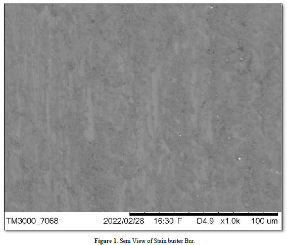
A Tabletop SEM (scanning electron microscope) is used for imaging and microscopic analysis of biological, inorganic, and man-made samples. It utilizes a focused beam of electrons to scan the surface of a sample to collect a 3D image of the sample. TM 3000 can be used for compositional imaging, stereoscopic observations with high resolution, and distance measurement [7]. The TM 3000 is interconnected with SwiftED 3000 for elemental mapping, acquiring spectra, and intensity profile of elements. The system is alternative option to optical microscopes, stereo microscopes, and confocal laser scanning microscopes [8]. In scanning electron microscopy teeth are evaluated in terms of qualitative surface roughness. The samples were mounted on metal stubs and middle 3rd portion of the crown was focused under the beam exerted by the SEM and examined under X1000 magnification.
Enamel Damage Index (EDI) [9] includes four scores:
Score 0 indicates smooth enamel surface without presence of scratches. Perikymata may be seen on enamel surface.
Score 1 indicates acceptable enamel surface with fine scattered scratches that involves 1-10% of enamel surface.
Score 2 indicates rough enamel surface with several coarse scratches or minor grooves that may involve 11-50% of enamel surface.
Score 3 indicates coarse scratches or wide grooves that may involve more than 50% enamel surface. Enamel damage in this score is visible with naked eye.
In the optical profilometer samples were mounted on metal stubs and middle 3rd portion of the crown was focused under the laser light and surface was observed on the screen.
Profilometer measurements include Ra, Rq, Rz, Rmax and Rt values and surface graphics. These values are:
Ra: Arithmetic average of Ra values in roughness profile
Rq: Geometric average of the deviations occurring in roughness profile
Rz: Average height of peak-to-valley
Rmax: Maximum roughness depth
Rt: Roughness depth
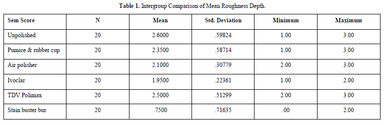
Intragroup and intergroup comparison was done to rule out for the mean roughness depth.
RESULT
The results obtained from Scanning electron microscopy study revealed that in case of unpolished surface the average score calculated is 3. In the pumice and rubber cup group average samples scored 3. In the group of air polisher average samples scored 2. In case of samples in ivoclar all the samples averaged a score of 2, the samples of TDV polimax averaged with score 3 and lastly in case of stain buster bur samples scores averaged with a 1.
The results obtained were statistically significant stating that the lowest mean value of 7500 in stain buster bur which means the lowest roughness value was seen in that group following ivoclar proxyt paste, air polisher, pumice and rubber cup, TDV polimax and lastly the group which was the unpolished group.
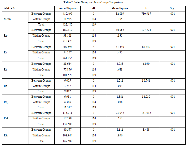
This analysis was done by Anova test followed by post hoc tukey test which showed the following result (Tables 1 & 2 and Figure 1).


Mean difference of values between group I and other respective groups showed a significant difference between group I and group VI showing value of 5.74 (values up to 2 decimal point), following the Rp and Rv value showing the most difference between group I and group VI along with group V showing value of 3.08 and 3.38 respectively (values up to 2 decimal point). The Stain buster group for Rsk sample showed highly significant difference with all the other groups except TDV polimax group having mean difference value of -.09 with pumice group. Again, the stain buster bur showed significant difference for Rkr value with all other group except TDV polimax.
Conclusively, the results obtained were highly in favor of the stain buster group both from Scanning electron microscopy as well as Optical profilometer.
This group showed the least hardness after polishing and revealed least loss of tooth structure which upon comparison with other groups was highly significant.
DISCUSSION
The objective of this study was to compare the efficacy of 5 different polishing systems on the basis of surface roughness it tends to create and also when it is compared with a non-polished surface. In our study, we did scaling with the help of ultrasonic devices because we wanted to evaluate the reduction of roughness caused post scaling by using different materials. In order to reduce the surface roughness after scaling process, various techniques and materials were used and all the materials which were used were of similar grain size.
The ideal requirements of a polishing pastes, good cleansing ability, minimal abrasion, and simultaneous polishing [1]. Hence all the materials that have been used were all capable of achieving the same.
Pumice is a light gray, highly siliceous material produced by volcanic activity used for polishing of tooth enamel, gold foil, dental amalgam and acrylic resins. The polishing application that was done by using rotary rubber cup was performed by the same researcher only by the weight of rotary instrument without extra pressure used with rotary rubber cup/brush. There is no standard in abrasiveness of paste among manufacturers [10].
In our study we did not get the results as the air polisher comparatively showed less surface roughness as compared to the pumice and rubber cup.
Today, the most widely used polishing material is rotary rubber cup, pumice or prophylaxis paste. This method often creates disappointment in settled colorations, it requires a long time and is tiring for the dentist so in order to be able to make the process faster and more efficient, the one of the devices developed is the air-flow polishing instrument ejecting compressed air, water and sodium bicarbonate that have its own advantages and disadvantages.
The Prophy-Jet is an excellent alternative instrument for removal of tooth stain and dental plaque [11] while we used air powder instrument in our study, the application was done the same way as done by some other researcher from 1-1.5 cm by approaching at a right angle to the tooth surface [10].
However, our study supports the argument that application by the air polisher is more effective option in reducing the surface roughness independent of the grain size, because prophylaxis paste that was used in paste application done by rotary rubber cup and the powder that was used in air-flow instrument were manufactured by the same manufacturer Proxyt paste by ivoclar is a fluoride rich prophylaxis paste. Proxyt fine and Proxyt coarse are available in the Proxyt Single Dose delivery form. The Proxyt Single Dose pastes have been developed with the aim of ensuring quick, easy and hygienic treatment of all patients. Proxyt fine without pumice is suitable for polishing tooth surfaces as well for cleaning top-quality restorations and implants. The paste is gentle to the gums and peri-implant tissue [12].
Similarly, in our study we were able to achieve a glossy surface visually and comparing it with the other pumice prophylaxis paste it showed statistically significant result, even when compared to air polisher it slightly showed a less of roughness depth.
Impregnated with an ultra-fine dehydrated abrasive polishing paste, activated with a few drops of water, Polimax provides an excellent polishing and natural shine on all surfaces.
In a study where TDV polimax and polished with Optimize (TDV) abrasive rubbers and Polimax Felt Disc (TDV) were used achieved a polished surface before placing a restoration. G Silva [13] in our study TDV polimax was unable to give desired result qualitatively and also quantitatively.
Stain buster burs were evaluated as the new material for polishing. These burs were made of glass fiber reinforced resins that were enriched by zircon and were designed for removing the colored layers, stain and cement from enamel surface. The surface characteristics of stain buster burs are abrasive power of fiber structure covering the entire work surface and divided into small fragments, when it contacts with a hard surface. While resin matrix is used, fibers occur; therefore, it also has the self-sharpening feature. It becomes sharp by itself, and the characteristics of abrasive are permanent. However, they slide over the tissues such as fibromatosis gingival membranes without cutting or trimming and they do not impact on soft tissue [13,16].
Although studies on the effect of burs on hard tooth tissues and especially surface roughness are not sufficient on the literature [14] for comparing the effects of bur on surface roughness, air-polishing method was preferred as it is known to leave rough surfaces and the prophylaxis paste which is the most commonly used polishing method in clinics [15]. In our study we observed the statistically significant result for stain buster bur when compared to the air polisher and pumice and rubber cup.
In the group used air-flow, a significant reduction could not be detected in the surface roughness hence stain buster bur may be an alternative method for traditional polishing material, because of providing the ease of application such as air polishing techniques and providing smooth surfaces like prophylaxis paste [15].
In this way, it was evaluated if the application of the products having the same abrasive properties be it rotary instruments or aerator devices it does affects the surface roughness. According to the statistical analysis of data, it was determined that reduction observed in roughness values of stain buster bur group has been significant when compared with the other groups but groups treated with air polisher and ivoclar proxyt paste where also able to show statistically significant result in terms of roughness and polishing.
CONCLUSION
Tooth polishing used to be a standard part of a dental cleaning appointment. The dentists use it to smoothen teeth so that plaque and bacteria which causes gingivitis, periodontitis or cavities do not stick to the tooth easily.
From the analysis of the results and within the limitations of the present study following conclusions can be drawn that our study tries to be a scientific guide for the clinical application of polishing processes. According to the results of our study, stain buster burs are seen as an alternative to traditional polishing materials, because it provides smooth surfaces like prophylaxis paste and ease of application like air-polishing technique. Stain buster bur showing the most impeccable result has 5 different bur shapes which can be used as per the area (e.g.: interdental area, cusps etc.) and can be used as alternative to traditional polishing materials, air polisher can also be a part of an alternative material as both of them created the least of surface roughness and has an ease of its application.
Future studies with more critically designed protocols, larger sample size and inclusion of various other biochemical and microbiological examination are necessary to further explore the potential of this perspective of periodontal treatment.
No Files Found


Internationally Accepted
Share Your Publication :